
A research group comprised of Professor Takeshi Matsui of the School of Bioscience and Biotechnology, Tokyo University of Technology, and Professor Hiroyuki Sasaki of the Rehabilitation Department, Tokyo Professional University of Health Sciences, has successfully developed a new staining methodology for electron microscopy.
Provided by Tokyo University of Technology
Uranyl acetate staining which is used to prepare biological samples for electron microscopy was originally reported in 1958, and since then, it has been used in electron microscopy facilities around the world for its simplicity and optimal staining effects. However, due to uranium being used as a material in weapons, international regulations on its use, acquisition, storage, and disposal are becoming increasingly restrictive, raising expectations for an alternative staining method for bioscience research.
In order to develop a safe and accessible alternative to uranyl acetate stain for electron microscopy using ultra-thin sectioning, the research group started by examining a variety of commercially available dyes used in optical microscopy. In the process, they learned that double staining with Mayer's Hematoxylin, a stain commonly used in optical microscopy, with a lead solution achieves a similar staining result to that of uranyl acetate combined with the lead solution for a variety of tissues and cells.
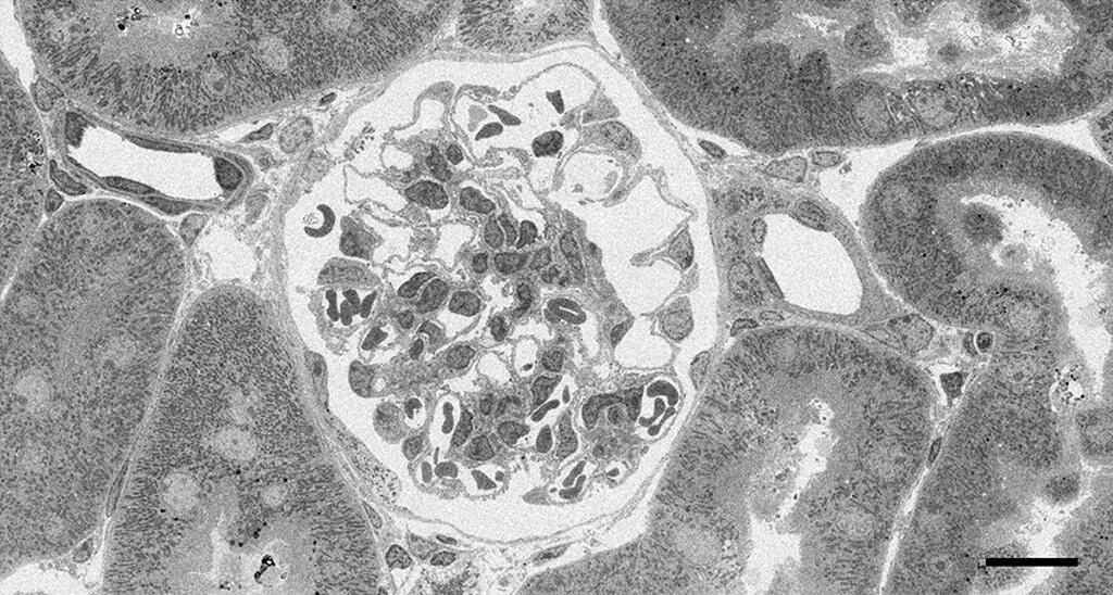
They also managed to stain a variety of organelles, such as nuclear chromatin, plasma membrane structures, ribosomes, glycogen, fat droplets, cell adhesion apparatus, and cytoskeletal systems, with a high degree of contrast using the Mayer's Hematoxylin method. The cell membranes in all samples were also stained particularly well. Backscattered electron images of 200-nanometer ultra-thin sections of mouse kidney observed with a field-emission high-resolution scanning electron microscope also showed that the Mayer's Hematoxylin and lead staining method provides wide-coverage, high-quality images of the renal cortical tubules and renal glomeruli.
Accordingly, the Mayer's Hematoxylin method is expected to serve as an effective replacement for the radioactive material uranyl acetate in terms of purchase, handling, use, storage, waste, and processing of the solution.
According to Professor Matsui, "We plan to further improve this new Mayer's Hematoxylin staining method and apply it various microscopy preparation steps, such as section staining as well as staining tissue sections before plastic embedding."
■ Mayer's Hematoxylin - lead staining: A useful staining method said to acidify the stain by adding citric acid and selectively stain the nucleus. It does not require separation using hydrochloric acid alcohol and does not suffer from the co-staining of the background.
Journal Information
Publication: Scientific Reports
Title: Novel electron microscopic staining method using traditional dye, hematoxylin
DOI:10.1038/s41598-022-11523-y
University Press release: https://www.teu.ac.jp/english/information/2022/0517.html
This article has been translated by JST with permission from The Science News Ltd.(https://sci-news.co.jp/). Unauthorized reproduction of the article and photographs is prohibited.