
A research group led by the National Institute for Basic Biology (NIBB) Division of Embryology graduate student Fumiko Matsukawa Usami and Professor Toshihiko Fujimori, in collaboration with Dr. Masatoshi Takeichi of RIKEN and Dr. Fadel Tissir of the Université Catholique de Louvain, Belgium, discovered that intercellular and intracellular cilia orientation within multiciliated cells of the oviduct is coordinated by two proteins, CELSR1 and CAMSAP3.
The oviduct, or fallopian tube, does not just transport the egg from the ovary in mammals, it is the organ that transports sperm cells and provides the environment for initial embryo development. In the oviduct, cilia (fine hair-like structures) orient in the same direction to carry the egg from the ovary to the uterus. The research group worked to discover the mechanism by which these cilia work across the epidermis as a whole.
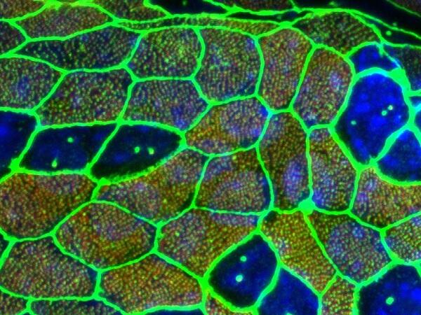
Super-resolution microscopy enables investigation of a wide expanse of cilia orientation. This was applied as a new way of establishing the direction of cilia within and between cells in investigating the cilia orientation of multiciliated cells.
The team found that in wild mice, both intracellular and intercellular cilia were orientated in the same direction. In a mouse with a CELSR1 mutation (CELSR1 mutant), the cilia orientation within cells was moderately maintained, although the intercellular coordination was disrupted. Furthermore, in the Ceslr1 mutant mouse, around 75% of cells showed an unusual microtubule enrichment near the cell boundary. In the CELSR1- deficient cells with enrichment of microtubules observed, cilia tended to orient in the direction of the microtubule enrichment.
What is more, with respect to the muticiliated cells in the oviduct of a mouse with a CAMSAP3 mutation (CAMSAP3 mutant), intracellular coordination of cilia was disrupted, with the direction random by cell.
Based on these factors, it was found that CELSR1 protein, which is involved with regulating cilial cell polarity, enriches the microtubules in the same direction across cells, coordinating the intercellular direction of cilia. It was also revealed that CAMSAP3, the microtubule binding protein, ensured that the cilia within each cell are oriented in the same direction.
Fujimori explains, "The epidermal cells of the oviduct are constantly exchanged over the course of a lifetime. Even if the oviduct is damaged, the cells are restored for the oviduct to regain its function. In future, we hope to find out how new cells are coordinated with their surrounding cells, and cell orientation is coordinated across the entire epidermis."
This article has been translated by JST with permission from The Science News Ltd.(https://sci-news.co.jp/). Unauthorized reproduction of the article and photographs is prohibited.