
A research group comprising Lecturer Masahiro Ohtsuka, Professor Shunsuke Muto and others at the Institute of Materials and Systems for Sustainability (IMaSS), Nagoya University, in collaboration with a research group comprising Dr. Makoto Tanaka and others at the Japan Fine Ceramics Center, has succeeded in using a transmission electron microscopic to precisely measure the position of a heterogeneous ion (dopant) that was added in a miniscule amount to an oxide ceramic, and has gone as far as clarifying the loopholes of the adjoining oxygen (the oxygen vacancy).
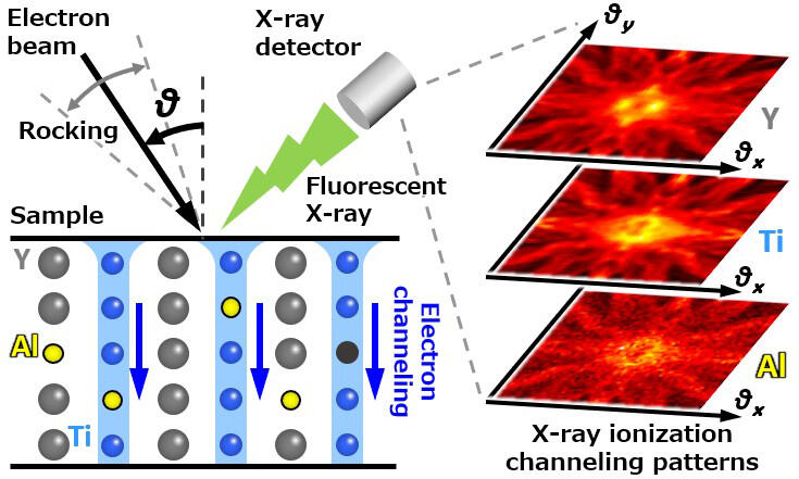
An oxide ceramic (yttrium titanate) is used as a protective membrane material in next-generation aircraft engines. When a miniscule amount of dopant (aluminum) is applied to it, the membrane structure is stabilized, and oxygen migration within the material is inhibited. Nevertheless, there is scant information regarding point defects such as dopant or vacancies scattered about without being arranged neatly within the material, and it is not easy to detect them even if measured using a large synchrotron radiation facility or the most advanced electron microscope.
With this in mind, the research group achieved the evaluation of information on minute defects using a laboratory-scale general-purpose electron microscope by combining a unique high-quality data acquisition method, a statistical data analysis method, and a precise theoretical calculation method. Specifically, in order to quantitatively determine the position and volume occupied by the aluminum of yttrium titanate (Y2Ti2O7), the group carried out analysis by means of the high angular resolution electron channeling X-ray spectroscopy (HARECXS) method, which utilizes the electron channeling effect that materializes in an electron beam incident on a crystalline sample in a transmission electron microscopic.
Credit: Nagoya University.
As a result of that analysis, the group was able to determine not only deformities in the dopant and the localized lattice in its vicinity, but also the position of very slight oxygen vacancies that only around 0.2% of the overall oxygen slips through.
Ohtsuka says that "In the future we plan to apply and develop our findings as a precise and high-throughput method for the point defects that govern the properties of functional materials such as semiconductors and dielectric and magnetic bodies, and also to expand it as a new analysis method for lattice defects that have conventionally been difficult to analyze, such as the random grain boundaries that are often seen in practical materials. We hope to promote materials development in a wide range of sectors through these advances."
■ Electron channeling effect: A phenomenon in which an electron beam incident on crystals branches into a number of Bloch waves (electron standing waves) as a result of sensing the periodic Coulomb potential of the crystals, and because their excitation probabilities change according to the incident angle (electron condition of refraction), the electron beam accentuates a different position and diffuses.
This article has been translated by JST with permission from The Science News Ltd.(https://sci-news.co.jp/). Unauthorized reproduction of the article and photographs is prohibited.