
This article was updated on August 30th, 2021.
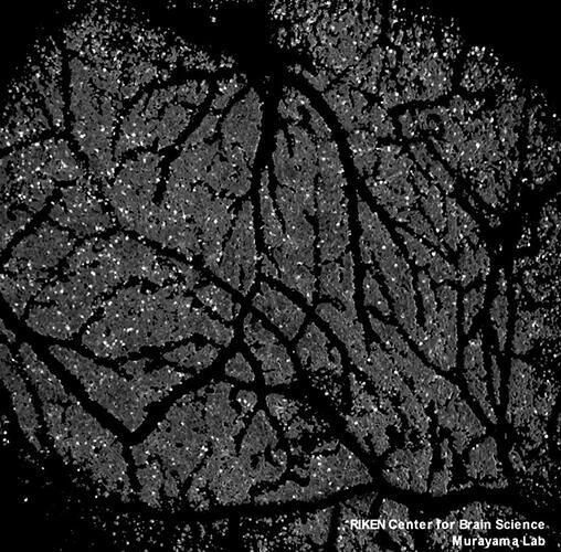
A joint research team led by Dr. Masanori Murayama of the RIKEN Center for Brain Science, including members from the University of Tokyo, Tokyo Institute of Technology, National Institute For Physiological Science, Juntendo University, Tohoku University, FOV, Nikon, and Hamamatsu Photonics, has developed the world's first two-photon microscope, the FASHIO-2PM (fast-scanning high optical invariant two-photon microscopy), featuring a wide field-of-view (FOV), high resolution, high speed imaging, high sensitivity, and practically no aberration. The team was able to successfully capture the activity of more than 16,000 neurons in the neocortex of a living mouse at a speed of 7.5 images per second from a single FOV, 36 times wider than a conventional microscope. According to team leader Murayama, "We can see even cortical layer 5 neurons of the mouse, and imaging from the wider FOV allows us to observe other brain regions simultaneously. This wide imaging allows us to observe the brain operating principles for perception, cognition, and decision making in the model animal for mental disorders in terms of the large network at the cellular level. We are proud to say that our microscope achieves all the critical parameters concurrently for wide FOV two-photon imaging, which has not been achieved in other microscopes." Their paper was published in Neuron and selected as journal cover art.
For the project, top experts in optics and calcium imaging came together as a nation-wide, all-star Japanese team to develop the large-scale aberration-free objective lens and the large-aperture, high-sensitivity, high-output photodetector for incorporation into the FASHIO-2PM. While the FOV of a typical two-photon microscope is currently 0.5 square millimeters, the FASHIO-2PM achieves 3 square millimeters, an area 36 times larger. The time resolution has also been greatly improved to 7.5 Hz. Some recent microscopes have increased the FOV at the cost of low resolution or slow recording speed, rendering them too slow to observe immediate intercellular cooperation, and as a result, functional structure at the cellular level across multiple regions remained unexplained. For this project, data from 15 brain regions within a single FOV was analyzed, and the research team was able to demonstrate, for the first time in the world, that the structure of the brain network was a small-world network rather than a scale-free network. According to Dr. Murayama, "In the future, we may be able to explain network dynamics at cellular resolution during perceptual decision tasks." The device will be released for sale by Nikon.
This article has been translated by JST with permission from The Science News Ltd.(https://sci-news.co.jp/). Unauthorized reproduction of the article and photographs is prohibited.