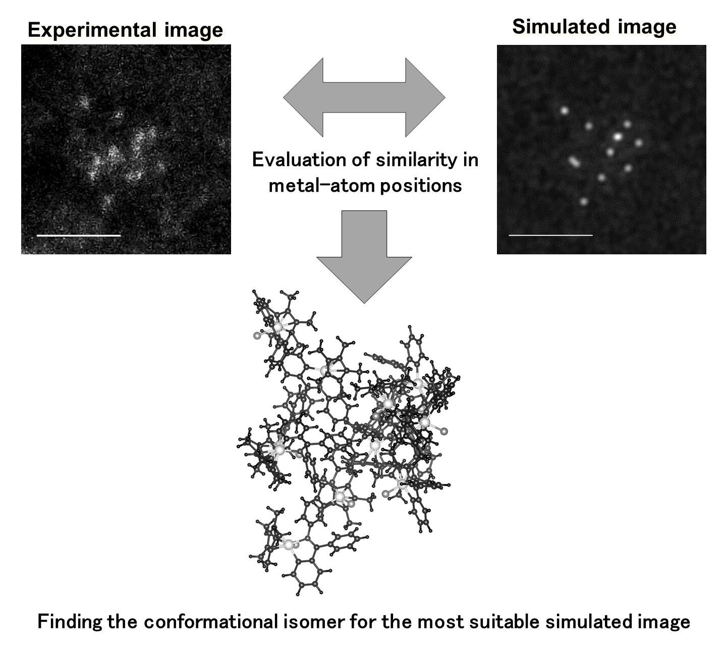
The research group led by Kenji Takada (at the time of research), Associate Professor Takane Imaoka, Assistant Professor Ken Albrecht (at the time of research), and Professor Kimihisa Yamamoto of the Institute of Innovative Research Laboratory for Chemistry and Life Science/ Hybrid Materials Unit, Tokyo Institute of Technology, has developed a method to determine the complex three-dimensional structure of macromolecules at the single-molecule level based on images obtained using an electron microscope with atomic resolution to determine the position of the metal atoms.
One current issue in the research world is that a structural analysis method for polynuclear metal complexes containing many metal atoms has not yet been established. Compounding this, a three-dimensional structure at the single molecule level has not been observed, even when using a scanning transmission electron microscope that is capable of observation at the single atom level. To tackle this issue, the research group reduced the electron beam intensity to a low acceleration voltage and low-current conditions to obtain electron microscope images without destroying the molecules that are vulnerable to electron beams. They also captured the images rapidly to shorten the irradiation time.
Dendrimers (dendritic polymer) fixed at a specific site on the iridium atom as marks were prepared for observation, and a metal immobilization reaction accompanied by cyclometalation was employed to synthesize and isolate iridium polynuclear complex molecules with 11-17 iridium atoms. In addition, the synthesized iridium polynuclear complex molecules were dispersed on graphene, and the carbon atoms were arranged in a hexagonal grid with a thickness of one atom. From there, high-angle annular dark-field scanning transmission electron microscopy (HAADF-STEM) was performed at a low acceleration voltage and with a low electron dose. The position of the iridium atom in each polynuclear complex molecule was then captured as a microscopic image. Furthermore, to clarify the three-dimensional structure of the original polynuclear complex from the microscopic image, theoretical simulations were performed.

Credit: Tokyo Institute of Technology.
Journal Information: Kenji Takada, Mari Morita, Takane Imaoka, Junko Kakinuma, Ken Albrecht, Kimihisa Yamamoto: Metal atom-guided conformational analysis of single polynuclear coordination molecules. https://www.science.org/doi/10.1126/sciadv.abd9887
As a result, it was possible to confirm the three-dimensional structure corresponding to the experimentally observed microscopic images of each polynuclear complex of iridium. It was then possible to proceed with the analysis of the three-dimensional structure efficiently. The agreement between the experimental and simulated images was evaluated based on the magnitude of the shift in the position of the iridium atom in the polynuclear complex in both images. The average deviation was approximately 0.3 nm with respect to the microscopic image of the three-dimensional structure to be analyzed, which is approximately equivalent to the size of one iridium atom; on top of this there was very limited error.
Associate Professor Imaoka stated, "In the future, the research group intends to further improve the electron microscopy observation and structural analysis methods developed in our study to analyze the three-dimensional structure of polymers with larger and more flexible structures and also materials with amorphous structures. The group also intends to determine the relationship between the structure and function of polymer materials."
This article has been translated by JST with permission from The Science News Ltd.(https://sci-news.co.jp/). Unauthorized reproduction of the article and photographs is prohibited.


