
A research group led by Associate Professor Eisuke Itakura of the Graduate School of Science, Chiba University, developed a stressed-mitochondria sensor, Mito-Pain, which helps label stressed mitochondria using fluorescent proteins. By performing an analysis using Mito-Pain and screening for compounds that can damage the mitochondria, it has become clear that functions of the causative genes for Parkinson's disease differ in response to specific types of mitochondrial stresses. These findings were published in Journal of Biological Chemistry.
Mitochondria are susceptible to damage by stresses such as reactive oxygen species. Accumulation of damaged, defective mitochondria adversely affects cells, leading to diseases such as Parkinson's disease, other neurological diseases, and cancer. Because of this, it is crucial to determine when defective mitochondria arise. However, methods that can detect specific stresses are limited.
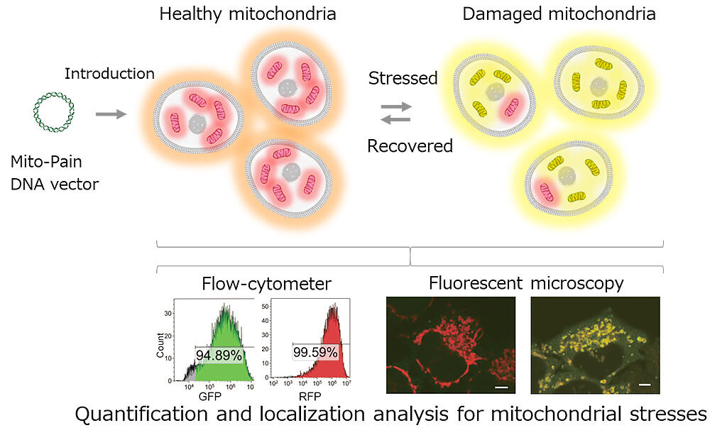
PINK1 is one of the causative genes for Parkinson's disease. Previous studies have shown that PINK1 is rapidly degraded in healthy cells; however, in defective mitochondria, it remains stabilized at the mitochondrial outer membrane. To exploit this property of PINK1 and leverage it as a marker for defective mitochondria, the research group generated Mito-Pain (DNA vectors) consisting of PINK1 and green fluorescent protein (GFP), T2A (self-splicing sequence), red fluorescent protein (RFP), and Omp25 (mitochondrial outer membrane protein). When Mito-Pain is introduced into cells, equal amounts of PINK1-GFP and RFP-Omp25 are produced from a single mRNA. From here RFP-Omp25 localizes to all mitochondrial membranes and PINK1-GFP is stably localized only on the membrane of defective mitochondria.
Healthy mitochondria were found to retain only RFP while defective mitochondria retained both RFP and GFP, thereby enabling quantitative analysis of an increased yellow (mixed red and green) signal as an indicator of an increase in the number of defective mitochondria. Fluorescence microscopy can also be used to analyze local mitochondrial stress through detection of defective mitochondria in the cell. When mitochondria become stressed and defective, PINK1 and other Parkin proteins, which are known to be products of the causative genes for Parkinson's disease, act together to degrade and remove defective mitochondria through autophagy (mitophagy). The research group investigated various compounds that cause mitochondrial stress using Mito-Pain. Depending on the type of compound, PINK1 alone may respond to mitochondrial stress, suggesting that PINK1 functions not only in mitophagy, but also in mitochondrial repair.
(Provided by Chiba University)
Associate Professor Itakura said, "It is thought that mitochondrial stress causes Parkinson's disease, neurological diseases, cancer, aging, etc. The details of mitochondrial stress can be studied in various diseases using Mito-Pain. I hope that the causes of the diseases can thus be elucidated."
This article has been translated by JST with permission from The Science News Ltd.(https://sci-news.co.jp/). Unauthorized reproduction of the article and photographs is prohibited.