
In addition to accurately capturing abnormal neural circuit activity, a research group consisting of Director Makoto Higuchi and Senior Researcher Masafumi Shimojo, both from the Department of Functional Brain Imaging, Institute for Quantum Medical Science, Quantum Life and Medical Science Directorate, and National Institutes for Quantum and Radiological Science and Technology (QST), and researchers from Scripps Research and Keio University School of Medicine, Department of Neurology, succeeded in developing an innovative technology that could image the earliest stages of the accumulation of dementia-causing substances in the brains of living animals.
Brain function in animals depends on the processing of information in circuits in which neural cells are intricately wired. Developmental abnormalities and functional impairments in specific circuits are the major causes of the cognitive impairments observed in many human patients with psychiatric and neurological disorders. However, it is technically difficult to visualize the structure and activity of these neural circuits. A further issue is that visualizing the earliest stage of pathogenic protein accumulation without damaging the animal brain has been impossible until now.
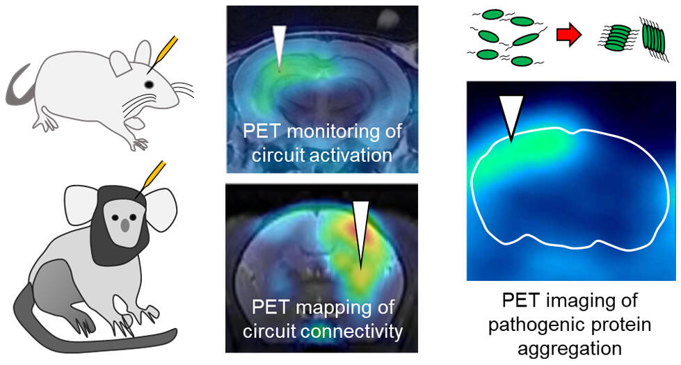
From this background, the research group introduced an intracerebral reporter, TMP, which does not naturally exist in animals, into cells that belong to a specific neuronal circuit. The researchers then intravenously injected an imaging agent that selectively binds to this intracerebral reporter. With this approach, they were the first to develop a technique for imaging circuit activity by positron emission tomography (PET). In addition, the reporter in the brain was divided into two parts. Each part was connected to a tau protein and expressed in the brain. This allowed the researchers to connect the two parts of the reporter in the brain only when the tau proteins remained connected, thereby allowing their imaging with PET. This allowed the precise visualization of the initial phase of tau protein accumulation, which has been challenging to detect until now.
Credit: QST
This technology will help capture various stages of pathological conditions, such as accurately detecting circuit connections and abnormal activity in the brain of disease model animals and promptly detecting the accumulation of pathogenic proteins that cause brain damage. It can also be used to visualize circuit hypoplasia as seen in animal models of brain development disorders. Furthermore, it may be applied to evaluate the effects of therapeutic agents on repairing circuit abnormalities and the effect of suppressing abnormal protein accumulation. The method is also expected to be useful for the development of therapeutic modalities for brain diseases.
According to Senior Researcher Shimojo, "This can be a tool for visualizing various stages of pathological conditions without damaging the animal brain, such as quickly detecting the accumulation of pathogenic proteins that cause brain damage. In the future, when this technology can be used, it will be possible to evaluate the effect of therapeutic agents for repairing circuit abnormalities and suppressing abnormal protein accumulation. I would also like to apply this technique for the development of therapeutic methods for brain diseases."
■ Intracerebral Reporter: A method of marking nerve cells using a harmless enzyme molecule called ecDHFR, which is not present in the brain of living mammals. This enzyme has the property of binding strongly to its unique antagonist, a compound called TMP. Therefore, a specific imaging agent that connects a fluorescent or radioactive reporter to the TMP can be administered to animals to allow visualization of specific neuronal cell circuits that express this enzyme via fluorescence microscopy or PET.
This article has been translated by JST with permission from The Science News Ltd.(https://sci-news.co.jp/). Unauthorized reproduction of the article and photographs is prohibited.