
A research group comprising Professors Hirosuke Shiura and Takashi Kohda of Yamanashi University, Fumitoshi Ishino of Tokyo Medical and Dental University and Professor Tomoko Kaneko-Ishino of Tokai University School of Medicine, among others, used a mouse model to demonstrate that the protease activity of the protein coded by the Peg10 gene, which is unique to mammals, is essential for maintenance of the vascular structure of the placenta. Peg10 was also found to play an important role in normal fetal growth during the second trimester through to late gestation of pregnancy.
Peg10 is an evolutionarily new gene acquired only by marsupials such as kangaroos and eutherians (a group of placental mammals) such as humans and mice. Studies by the group have shown that this gene is derived from retrotransposons or retroviruses and is essential for early placental development, which is vital for fetal development and growth during pregnancy in eutherians. These findings suggest that a virus (or virus-like element) that infected the common ancestor of mammals became a new gene, Peg10, and contributed significantly to the evolution of a mammalian-specific mode of development, namely fetal life, where mothers incubate their offspring in their uterus.
The research group examined mice lacking the protease activity of the Peg10 protein. The protease-deficient mice did not display abnormal placental formation or lethality in the early embryonic period, which was observed in Peg10-deficient mice. Furthermore, placental and fetal growth was poor after the second trimester, and most individuals died perinatally. As the placenta is an important tissue that supports fetal growth, and poor placental growth often precedes fetal abnormalities, the poor fetal growth and perinatal lethality observed in these mice may be caused by placental insufficiency.
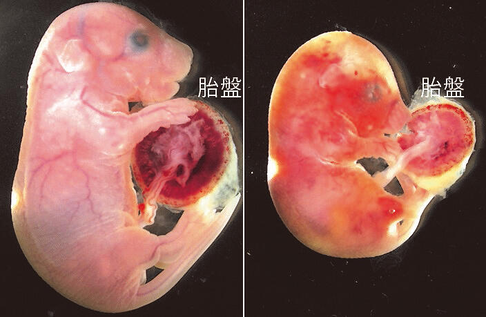
Provided by Hirosuke Shiura (University of Yamanashi) and Fumitoshi Ishino (Tokyo Medical and Dental University (TMDU))
Detailed examination of the placental status revealed severe damage to the fetal capillaries in the placenta leading to the fetus. The supply of oxygen and nutrients required for fetal growth and excretion of unwanted metabolites are carried out between fetal blood flowing through the fetal capillary and the maternal blood that fills the placenta outside it. In the architecture of this feto-maternal interface, Peg10 was found to be expressed in trophoblast cells (cells present only in embryonic mammals) that surround fetal capillaries and are in direct contact with maternal blood.
"This time, we have shown that Peg10 functions to maintain placental vascular architecture and has essential roles in normal fetal growth after the second trimester of pregnancy," said Dr. Shiura. "For the establishment of a fetus in mammals, solving the issues of creating a new organ called the placenta and evasion of the foreign fetus and placenta from the maternal immune system is necessary. We wish to proceed with the study of Peg10 as a key gene."
■ Protease: A general term for enzymes that cleave and break down proteins. The retrotransposon retrovirus that eventually became Peg10 cleaves its own protein through this activity and synthesizes multiple functional proteins. Peg10 inherited this functionality and can cleave itself.
This article has been translated by JST with permission from The Science News Ltd.(https://sci-news.co.jp/). Unauthorized reproduction of the article and photographs is prohibited.