In collaboration with the RIKEN Center for Brain Science and the University of St. Andrews in the United Kingdom, the Research Group of JSPS Fellow Xiangsunze Zeng, graduate student Yuko Komanome, and Professor Akinao Nose, all from the Graduate School of Frontier Sciences at the University of Tokyo, showed for the first time that the sensory feedback from exercise experience is essential for the development of motor circuits by specifically inhibiting the function of nerve cells responsible for somatosensory feedback in Drosophila by genetic manipulation.
Much like a human baby moves its hands and feet inside its mother's body, animals also begin to move while in the uterus or egg. One hypothesis suggests that these spontaneous and immature activities in the early stages of motor development are essential for the development of appropriate neural circuits by moving muscles on a trial basis and learning the results through somatosensory feedback. However, this hypothesis was yet to be verified. It has also been observed that during development of the cranial nerve system, a primitive circuit is first constructed by electrical synapses. This primitive circuit that produces spontaneous activity is then organized into a mature circuit that produces coordinated activity as development progresses. However, its significance was not well understood.
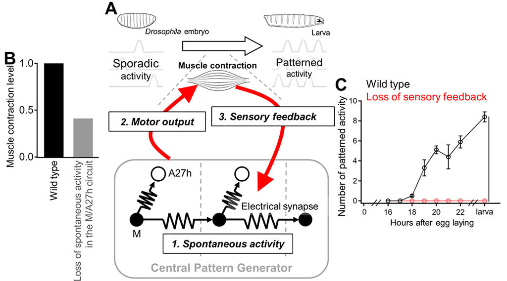
The research group used Drosophila embryos and larvae as models. They observed the transition of the activity pattern of the central pattern generation circuit (CPG) using a technique called calcium imaging, which visualizes neural activity. The results showed that only sporadic activity occurs within the central nervous system during the early phase of circuit development. However, as development progresses, some ganglia begin to synchronize and gradually show waves of regulated activity that propagate throughout the ganglion, as seen in post-hatching larvae.
To investigate the role of this somatosensory feedback activated by motor output during development, they utilized mutants with specifically inhibited somatosensation and examined the activity patterns of CPGs. They found that stereotyped neural activity corresponding to motor patterns did not occur at all in the mutants. Furthermore, through cell-specific calcium imaging, electrophysiology, and activity inhibition studies, the researchers identified a specific kind of electrical synapse-dependent neural circuitry, M/A27h, in which sensory feedback operates and serves as a nucleus for CPG formation. This neural circuit consists of two pairs of neurons (A27h and M cells) that are present in each ganglion of the spinal cord (abdominal ganglion in Drosophila). The absence of sensory feedback leads to the absence of electrical synapses within the M/A27h circuit and, as a result, the absence of neural activity, indicating that functional expression of this circuit depends on motor experience. Inhibition of spontaneous activity and electrical synapses within the M/A27h circuit led to severe impairments in larval spontaneous movements, indicating that the activity of this circuit plays an essential role in shaping motor circuits.
"We are finding molecules and cellular mechanisms that are conserved across species," Professor Nose stated, adding, "In the future, we hope to clarify the basic principles of motor circuit development, which self-organizes through feedback from immature outputs."

This article has been translated by JST with permission from The Science News Ltd.(https://sci-news.co.jp/). Unauthorized reproduction of the article and photographs is prohibited.


