
The phospholipids (PLs) that make up biological membranes that are susceptible to oxidation, and oxidized phospholipids (oxidized PLs) are readily generated. It is becoming clear that oxidized PLs affect various physiological activities, such as cell death and control of inflammatory responses, and are involved in the development of various diseases. However, due to the complicated mechanism of PL oxide formation, the number of PL oxides whose structures have been identified so far is extremely small. What kind of molecular species are actually increased or decreased at the onset of disease, and the nature of their localization, was completely unknown.
A research group has developed a comprehensive structural library of oxidized phospholipids (a total of 465 types of oxidized PL structures), which are the causative agents of various diseases. The group has also developed visualization technology. This research group consists of Assistant Professor Matsuoka Yuta, Professor Ken-ichi Yamada, Professor Kazuhiro Nishiyama, and Professor Motohiro Nishida, all of the Kyushu University Faculty of Pharmaceutical Sciences, as well as Associate Professor Masatomo Takahashi, Associate Professor Yoshihiro Izumi, and Professor Takeshi Bamba, all of the Kyushu University Medical Institute of Bioregulation, and finally Assistant Professor Yuki Sugiura and Professor Makoto Suematsu, both from the Keio University School of Medicine. Application of the structural library produced by this team revealed that as many as 70 types of oxidized PL are produced in the liver tissue in acetaminophen-induced acute liver injury model mice. Furthermore, a new heavy oxygen (18O2) -labeled mass spectrometric imaging method was developed for visualizing oxidized PL, and it was found that oxidized PL was accumulated at the damaged site of the liver tissue. These findings are expected to contribute to the discovery of new bioactive lipids, clarification of pathogenic mechanisms of oxidative stress diseases, and biomarker exploration. The group's findings were published in the online version of Nature Communications.
Oxidized PLs are believed to be involved in the development of various diseases such as liver and cardiovascular diseases and can have several effects, including controlling the immune response of each living tissue and inducing ferroptosis, which is a cell death mechanism newly proposed in recent years. However, oxidized PLs are generated by complex lipid peroxidation reactions, and therefore, several oxidized PLs are believed to be present at the onset of disease. However, until now, only a few molecular species have been evaluated in oxidized PL studies. This is because the number of structurally identified oxidized PLs is very low. As a result, it was not entirely clear which oxidative PLs actually increase or decrease at the onset of disease, or the site at which they are generated within biological tissues.
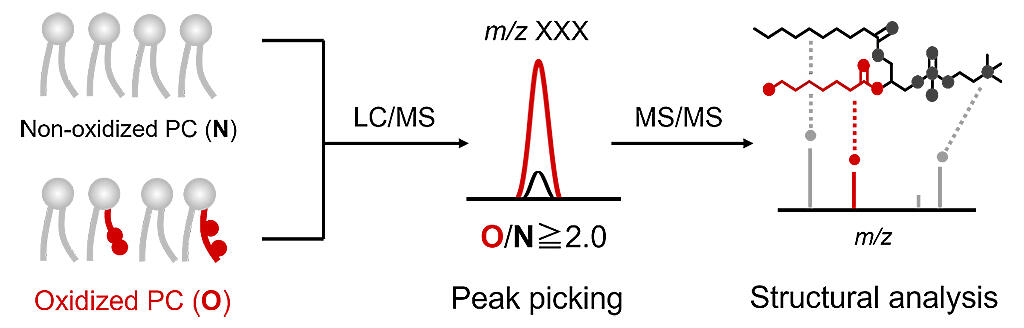
The research group first utilized non-target analysis using liquid chromatography-mass spectrometry (LC / MS / MS) as a technique for searching for unknown oxidized PLs. They constructed a structural library of phosphatidylcholine (PC) -derived oxides (oxidized PCs), which have the highest abundance in the body among PLs. When specimen PCs were oxidized in vitro, MS peaks with increased intensities were comprehensively analyzed. A total of 465 oxidized PC structures were successfully identified, and these were listed in a structural library. Using this structural library, the researchers comprehensively analyzed the oxidized PCs occurring in acetaminophen-induced acute liver injury model mice. While it has been reported that lipid peroxidation is involved in the progression of this disease, the oxidized lipids generated are unclear.
As a result of the analysis, the researchers found that 70 types of oxidized PCs increased in abundance when liver damage occurred. Furthermore, it was found that among oxidized PCs, the amount of PC dioxide with an epoxy group / hydroxyl group increased remarkably prior to liver damage. Next, to investigate the site in the liver where tissue PC dioxide is produced, an oxidized PC mass spectrometric imaging method using a heavy oxygen (18O2) label was developed.
Provided by Kyushu University
In this study, mice were given heavy oxygen to inhale, meaning that in vivo oxidized PC became labeled with that heavy oxygen. After that, mass spectrometric imaging of heavy oxygen-labeled-oxidized PC was performed, enabling more sensitive and selective imaging. By using this method, it was discovered that PC dioxides are accumulated at damaged sites (CYP2E1 expression sites) in the liver tissue. Professor Yamada said, "The analysis of oxidized PLs, which have complicated chemical structures, was a daunting task, but we were finally able to put our findings together into a paper. By applying this analytic technology, we would like to gain a deeper understanding of the diversity of oxidized lipids and their significance in living organisms."
This article has been translated by JST with permission from The Science News Ltd.(https://sci-news.co.jp/). Unauthorized reproduction of the article and photographs is prohibited.