A research group led by lecturer Koji Kikuchi of the Department of Molecular Pharmacology, Faculty of Life Sciences, Kumamoto University (at the time of research), Professor Kei-ichiro Ishiguro of the Department of Chromosome Biology, Institute of Molecular Embryology and Genetics, Kumamoto University, Professor Kenji Shimamura of the Department of Brain Morphogenesis, Kumamoto University, and visiting professor Shin-ichi Hisanaga of the Molecular Neuroscience Lab, Department of Biological Sciences, Tokyo Metropolitan University, has successfully identified a new regulatory system that controls neuronal morphology and motion.
It is known that when a cell functions, it must assume a form suitable for the expression of its function, and that this form is controlled by the cytoskeleton. Microtubules, a part of the cytoskeleton, have the property of repeating dynamic stretching and contraction due to the binding and separation (polymerization and depolymerization) of tubulin, a protein that makes up the cytoskeleton. Such microtubule dynamics are essential for the expression of cellular functions that involve changes in morphology, such as cell motility. While various molecules involved in microtubule dynamics have been identified, their regulatory mechanisms remain unclear because of the diversity in cellular functions.
The research group focused on the MAP7 family of microtubule-binding proteins and analyzed the function of Map7D2, which belongs to this family. Their results revealed that Map7D2 regulates the morphology and movement of cultured cells in the nervous system by binding to and directly stabilizing microtubules. Next, they conducted a functional analysis of Map7D1, another member of the MAP7 family, and found that Map7D1 stabilizes microtubules and regulates their morphology and movement through the maintenance of tubulin acetylation, which is involved in microtubule stabilization.
Despite belonging to the MAP7 family, Map7D2 and Map7D1 stabilized microtubules and regulated their morphology and movement by different mechanisms, indicating the existence of functional diversity among the family members.
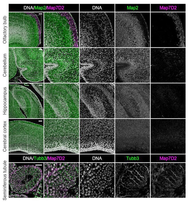
In addition, the research group analyzed the expression distribution of Map7D2 in different organs in rodents. Map7D2 is specifically expressed in the brain and testis, with particularly high expression in the olfactory bulb and glomerular layer where the axons of olfactory neurons accumulate in the brain, and in Sertoli cells in the testicular duct. Analysis of the expression distribution suggests that Map7D2 may regulate neuronal morphology by regulating microtubule stabilization in neurons in vivo.

Provided by Kumamoto University
"We initially assumed that Map7D1 and Map7D2, which are our key research targets, would have similar functions because they belong to the same family, but it became clear that, in fact, there is actually functional diversity among the family and that different mechanisms stabilized microtubules," commented Dr. Kikuchi. "Moving forward, we would like to use mouse models to understand the physiological significance in vivo, and in the future, we would like to shed light on the relationship with pathological conditions such as neurological diseases."
This article has been translated by JST with permission from The Science News Ltd.(https://sci-news.co.jp/). Unauthorized reproduction of the article and photographs is prohibited.


