
The joint research group of Assistant Professor Sho Kurihara, Assistant Professor Masayoshi Tei, and Professor Hiromi Kojima of the Department of Otorhinolaryngology, Jikei University School of Medicine, Professor Hirotaka James Okano of the Division of Regenerative Medicine, Jikei University School of Medicine, Associate Professor Junichi Hata of the Graduate School of Health Sciences, Tokyo Metropolitan University, and Professor Masato Fujioka of the Kitasato University School of Medicine, announced that they had succeeded in visualizing the three-dimensional structure of the olfactory nerve. By combining 9.4T ultra-high field MRI and nerve tractography (a 3D modeling technique to visually represent nerve tracts), the research group revealed the projection of the olfactory nerve from the nasal cavity to the central nervous system while maintaining the vertical and anterior-posterior axis, as well as their precise distribution. The result is expected to lead to the development of an objective evaluation method for olfactory disorders. The results were published in the September 6 issue of the international scientific journal Communications Biology.
Provided by the Jikei University School of Medicine
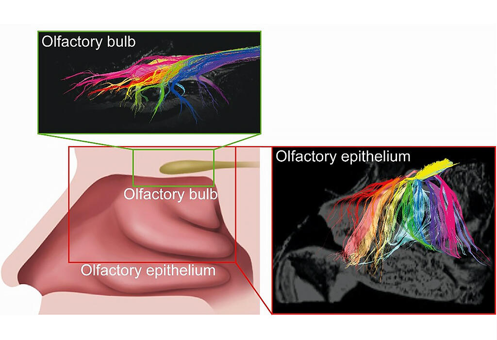
The sense of smell functions through the adhesion of volatile odor molecules to olfactory nerve cells in the nasal cavity and the transmission of nerve signals to the cerebrum via the primary olfactory center (olfactory bulb). Olfactory disorders are caused by upper respiratory tract infections, chronic sinus infections, and trauma, etc., and also appear as a precursor symptom of neurological diseases such as dementia and Parkinson's disease. In addition to reducing patients' quality of life, it has also been reported to lead to cognitive decline and shortened life expectancy, necessitating early detection and treatment. The risk increases with age, and the incidence rate is approximately 70% for those over 80 years of age. On the other hand, subjective symptoms are low, and all of the multiple types of tests are based on the patient's subjectivity, so there is a need for an objective evaluation method.
In this study, the research group investigated the possibility of visualizing the olfactory nerve by applying nerve tractography, one of the MRI analysis techniques already in clinical use in neurosurgery, to the nasal cavity. Nerve tractography can visualize nerve fibers by using diffusion-weighted images that capture the direction and speed of the movement of water molecules.
When the technique was first applied to mice, the same patterns of nerve fiber projection were visualized as those that were previously reported using neural tracers in genetically engineered mice. It allowed researchers to confirm that the nerve fibers distributed in the complex structure of the mouse nasal cavity project from the olfactory epithelium to the olfactory bulb, and how they project while maintaining the anterior-posterior and vertical axes.
Next, the same technique was applied to the common marmoset, a primate with a nasal cavity morphology similar to that of humans.
Nerve fibers projected while maintaining the anterior-posterior axis and the vertical axis were visualized as in mice. Then, immunohistological analysis was performed to verify whether the visualized nerve fibers were olfactory nerves. Histological sections of the nasal cavity were prepared, immunostained for olfactory nerve markers (OMPs), and analyzed by overlaying nerve tractography images of the same anatomic sites.
The results showed that the distribution of nerve fibers coincided with the staining sites, indicating that the olfactory nerve was depicted by nerve tractography. On the other hand, the nerve tractography did not recognize thin olfactory nerves, indicating that the olfactory nerves may be distributed over a wider area than was previously thought.
Based on these results, the technology was applied to humans by using donor bodies. The olfactory nerves were distributed similarly to as in mice and common marmosets. Further imaging with a cryoprobe revealed that the nerve fibers run in the olfactory bulb while maintaining their vertical and anterior-posterior axes. Combined with data from normal imaging, the researchers were able to create an olfactory nerve map that showed from which region of the nasal cavity the nerve fibers projected to the olfactory bulb.
Furthermore, when the technique was applied to a donor body with a nasal sinus surgery scar, the olfactory nerve was found to be shorter than in a donor body without the surgery scar. Although the statistical significance of this difference is unclear because the sample size was only a single donor body, the results indicate that nerve tractography can be used as an objective method to evaluate the olfactory nerve.
Kurihara said, "The MRI we used in our research is an experimental machine, so we will verify optimal imaging conditions with a clinical MRI that is capable of imaging people. We would also like to proceed with analyzing olfactory neuropathy models in animal experiments. Through these studies, we hope to develop an objective method that uses senses other than smell to test for olfactory neuropathy."
Journal Information
Publication: Communications Biology
Title: MRI tractography reveals the human olfactory nerve map connecting the olfactory epithelium and olfactory bulb
DOI: 10.1038/s42003-022-03794-y
This article has been translated by JST with permission from The Science News Ltd.(https://sci-news.co.jp/). Unauthorized reproduction of the article and photographs is prohibited.