
Cells have numerous organelles that coordinate to support various vital intracellular processes. In plant cells, photorespiration associated with photosynthetic reactions is performed via three organelles: chloroplasts, peroxisomes, and mitochondria. Peroxisomes and mitochondria have long been observed to come into physical contact with chloroplasts, a trait believed to be related to photorespiration. However, the way the shape and positioning of the organelles changed in response to light remained poorly understood.
A team including Postdoctoral Researcher Keiko Midorikawa of the RIKEN Center for Sustainable Resource Science (currently Project Research Associate at the Center for Bioscience Research and Education, Utsunomiya University), Visiting Senior Researcher Yutaka Kodama (Professor at the Center for Bioscience Research and Education, Utsunomiya University), and Team Leader Keiji Numata, (Professor at the Graduate School of Engineering, Kyoto University) came together to shed light on this process. The team used electron microscopy images and 3D image reconstruction techniques to reproduce Arabidopsis mesophyll cells as high-resolution 3D images. The team also quantitatively showed, for the first time, that the intracellular organelle morphology and contact areas between organelles were different between light-exposed cells (light treatment group) and those that were not light-exposed (dark treatment group). The results were published in the online edition of PNAS Nexus.
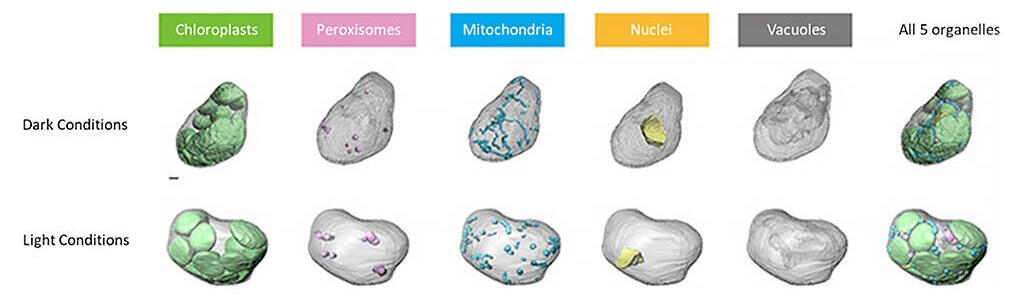
Arabidopsis thaliana phloem cells containing five organelles: chloroplasts (green), peroxisomes (pink), mitochondria (light blue), nuclei (yellow), and vacuole (gray), reconstructed in 3D using field emission scanning electron microscopy (FE-SEM). Three cells each were selected from dark-treated and light-treated leaves, for a total of six cells analyzed. The image shows one of the cells belonging to the dark- or light-treated groups analyzed, respectively.
Provided by RIKEN
The research team used array tomography by field emission scanning electron microscopy (FE-SEM). This array tomography method allows observations with a wide field of view while maintaining high resolution. Using this technique, the team successfully reconstructed an Arabidopsis mesophyll cell as a 3D image.
Using this image, the group compared the volume and sphericity of the three organelles between the light and dark treatment groups. The results showed that the light-treated group contained more large-volume peroxisomes. Mitochondrial volume did not differ between the two groups, but the light-treated group had a higher sphericity, that is, more mitochondria with simpler morphology.
The group analyzed chloroplast, peroxisome, and mitochondrial contact frequency (number of contact sites) and contact areas. Contact sites and contact areas were determined by algorithms provided by the image analysis software Imaris. A comparison of contact frequency, total contact area per cell, and area per contact between the groups revealed that the contact frequency tended to be higher in the light treatment group. Of note area per contact was significantly larger. These results suggest cellular photoreception encourages cells to expand existing contact sites rather than increase new contact sites between organelles.
When cells are exposed to light, photorespiration becomes active in line with photosynthetic reactions. As revealed in this study, the changes in peroxisome and mitochondrial morphology and the expansion of the chloroplast-peroxisome-mitochondria contact areas are thought to promote more efficient photorespiration.
In the future, obtaining details of organelles under the various stress conditions (not just light and dark) to which plants are exposed could lead to the development of a method to quantitatively evaluate cell metabolic efficiency.
Journal Information
Publication: PNAS Nexus
Title: Three-dimensional nanoscale analysis of light-dependent organelle changes in Arabidopsis mesophyll cells
DOI: 10.1093/pnasnexus/pgac225
This article has been translated by JST with permission from The Science News Ltd.(https://sci-news.co.jp/). Unauthorized reproduction of the article and photographs is prohibited.