
In the brain, synapse size changes with memory and learning, which regulates the strength of signals traveling through nerves, but the details of the mechanisms that regulate synaptic structure are not well understood. The research group of Researcher (at the time of the research) Yosuke M. Morizawa and Professor Ko Matsui of the Graduate School of Life Sciences, Tohoku University, discovered that cerebellar Bergmann glial cells phagocytose (a cellular process in which phagocyte cells ingest/engulf other cells or particles) synaptic cells, and showed that learning enhances this synaptic engulfment and that inhibiting the phenomenon reduces the learning effect. Matsui said, "The same thing is thought to occur in the cerebral cortex. In humans, synapses are pruned from childhood to adolescence, and if there is insufficient pruning then the result is autistic spectrum disorder, but if excessive pruning occurs, it results in schizophrenia. Focusing on glial cells may lead to the development of new treatment methods." The group's findings were published in Nature Neuroscience.
Provided by Yosuke M. Morizawa, Ko Matsui, Tohoku University.
Horizontal optokinetic response (HOKR) is a function that adjusts the image on the retina as much as possible by involuntarily moving a person's eyes according to the scenery, such as when looking out of a train window. Since HOKR learning is known to take place in specific areas of the cerebellum, the research group focused on this and cerebellar changes, particularly on phagocytosis and synapses.
To visualize phagocytosis, the researchers created a new mouse with genetic mutations that express a red fluorescent protein (pHRed), which is not easily degraded in lysosomes, only in target neurons at specific time points, and established a method to track fragments in neurons that were taken up by glial cells. In addition, they constructed brain tissue in three dimensions by thinly scraping the surface of a block of brain tissue with an FIB and continuously observing the surface of the blocked tissue with a scanning electron microscope.
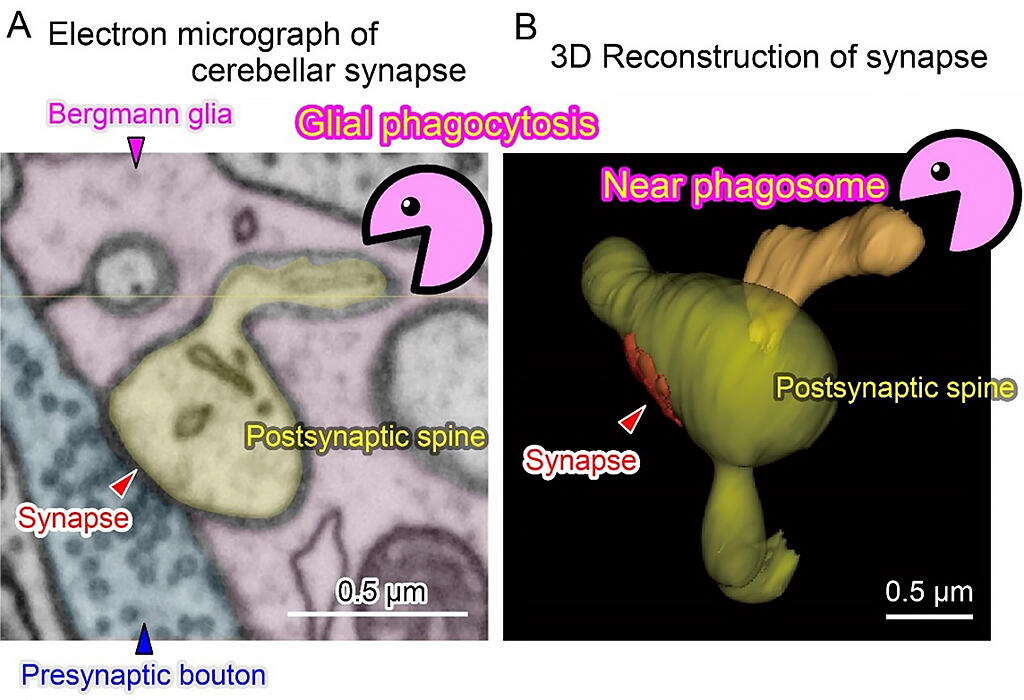
By utilizing these techniques, they succeeded in observing in detail the process by which Bergmann glial cells partially incorporate synaptic and non-synaptic neuronal structures. For example, the researchers obtained a variety of images of the process, including the moment when Bergmann glia cells are about to phagocytose a synaptic site, and immediately after the phagocytosis is completed and the original nerve site is completely disconnected. Although phagocytosis by glial cells has been reported in brain injuries and in the very early stages of growth and development, this is the first time that the critical moment of phagocytosis of spines by glial cells in the healthy brain has been captured in detail with an electron microscope.
When vertical stripes were repeatedly moved left and right in front of the mice's eyes, the amplitude of their eye movements increased due to HOKR learning. After the training, phagocytic activity was activated four times every hour, and the volume of the spines was reduced after 24 hours. On the other hand, administration of phagocytosis inhibitors suppressed the decrease in dendritic spine size and partially inhibited HOKR learning. Furthermore, a gene important for phagocytosis of Bergmann glial cells was found, and a portion of HOKR learning was also inhibited in mice that were specifically deficient in the gene.
Journal Information
Publication: Nature Neuroscience
Title: Synaptic pruning through glial synapse engulfment upon motor learning
DOI: 10.1038/s41593-022-01184-5
This article has been translated by JST with permission from The Science News Ltd.(https://sci-news.co.jp/). Unauthorized reproduction of the article and photographs is prohibited.