
The research group of Professor Kousei Ishigami and Assistant Professor Yuzo Yamasaki of the Department of Clinical Radiology, Graduate School of Medical Sciences, Kyushu University, Lecturer Abe Kohtaro from the Department of Cardiovascular Medicine, Kyushu University, and Clinical Development Department Manager Takenori Fukumoto from Konica Minolta, has developed the world's first system to diagnose pulmonary thromboembolisms by evaluating abnormalities in blood flow distribution that are suggestive of the disease. These inferences are based on changes over time in the X-ray permeability of the lungs in serial X-ray images, and by determining abnormal lung region findings in serial X-ray images or simple chest X-rays that are taken at the same time.
Provided by Kyushu University
Pulmonary hypertension is a group of progressive diseases with a poor prognosis in which blood pressure in the pulmonary arteries (the blood vessels that carry blood from the heart to the lungs) is elevated, resulting in heart and lung dysfunction. Chronic thromboembolic pulmonary hypertension (CTEPH) is a member of this group, in which the persistent formation of blood clots in the pulmonary arteries causes pulmonary hypertension due to widespread obstruction of pulmonary blood flow. It is a rare disease with approximately 4,600 patients in Japan, but this number is on the rise.
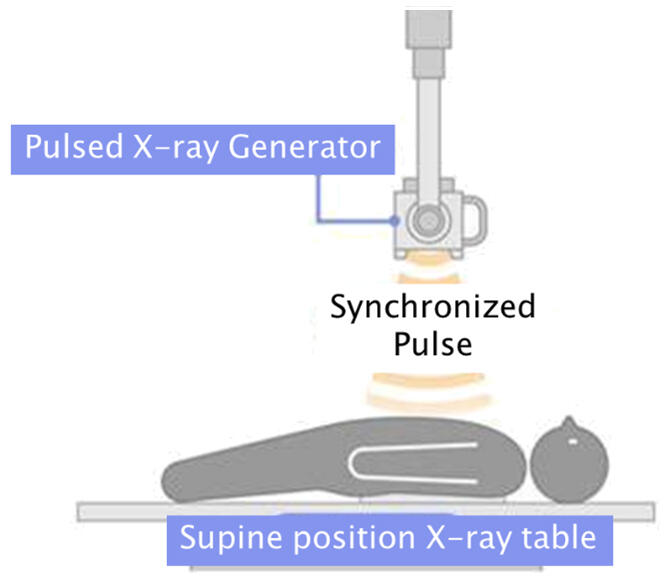
Dynamic chest radiography (DCR) is a technique that uses the same equipment as a simple X-ray to take a series of X-ray images over a seven to 10-second interval in which the patient is holding their breath, which allows for the acquisition of about 15 frames/second of continuous X-ray images. This technique does not use contrast agents or radionuclides, and the radiation dose is lower than the International Atomic Energy Agency's standard for frontal and lateral chest X-ray, about one-tenth the radiation dose of pulmonary ventilation and blood flow scintigraphy (the use of a radioactive tracer in a patient's blood).
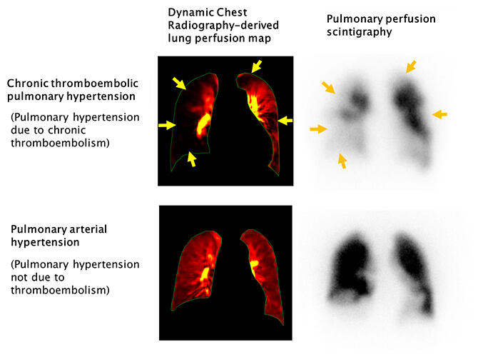
The research group developed a system to diagnose pulmonary thromboembolisms by evaluating abnormalities in blood flow distribution that were suggestive of a pulmonary thromboembolism based on changes over time in the X-ray permeability within the lungs on the series of X-ray images that they obtained, and this information was then combined with a simple X-ray or abnormal findings from the lung region in a series of original X-rays. The system's usefulness in detecting CTEPH was verified by a radiologist reading of data from 50 existing cases of pulmonary hypertension.
The results showed that the system has a high diagnostic performance, with a sensitivity of 97%, a specificity of 86%, and a diagnostic accuracy of 92%, and high agreement with pulmonary ventilation and blood flow scintigraphy, which is specified as the first choice for diagnosis in the guidelines (agreement rate of 90%). This is the first demonstration of a chest X-ray dynamic imaging system as a new diagnostic method for CTEPH, and it does not use contrast agents or radionuclides, is easy to use, and has lower radiation exposure. Kyushu University and Konica Minolta have jointly applied for a patent for the system they developed, and the application is currently pending.
Provided by Kyushu University
Yamasaki stated that, "We were able to create an organization in which diagnostic imaging physicians, cardiologists, and companies could cooperate with each other, and we believe that this led to our achievements. In the future, we hope to establish solid evidence through multicenter clinical trials and to apply the system to acute pulmonary thromboembolism. We would also like to use artificial intelligence to establish a system that even allows non-specialists to make decisions."
This article has been translated by JST with permission from The Science News Ltd. (https://sci-news.co.jp/). Unauthorized reproduction of the article and photographs is prohibited.