
A research group led by Assistant Professor Masashi Tanimoto and Professor Shin-ichi Higashijima of the Neuronal Networks Research Group, Exploratory Research Center on Life and Living Systems, National Institutes for Natural Sciences (National Institute for Basic Biology, NIBB), has fabricated a tiltable objective microscope that allows imaging while the biological specimen and objective lens are tilted and vibrated together. They used this microscope to successfully visualize the activity of hair cells and vestibular ganglion neurons in the otolith organ of zebrafish larvae by in vivo calcium (Ca2+) imaging, which is suitable for in vivo observation.
Provided by NIBB
Human head movements are thought to be received by the hair cells in the vestibular system of the inner ear. However, it is extremely difficult to measure the activity of hair cells during head movement, and how individual cells are active in vivo during head movement had not been examined.
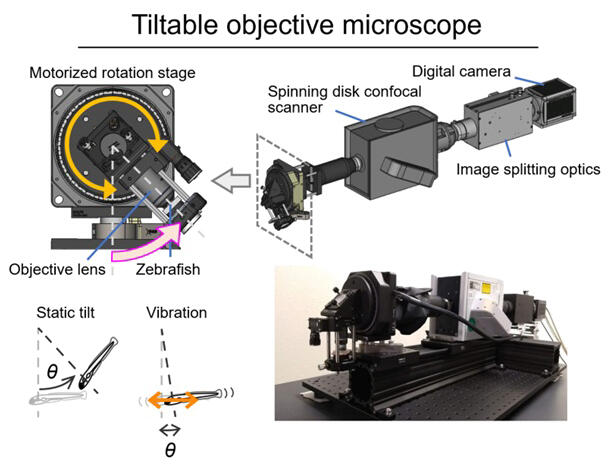
In response to this, the research group designed and assembled a tiltable objective microscope that combines a motorized rotation stage, a spinning disc confocal scanner and a camera for imaging biological samples while tilting and vibrating them. The motorized rotation stage allows imaging while the biological sample and objective lens move together. Slow, large-angle rotation produces a static tilt stimulus in the biological specimen, while quick, small-angle reciprocation produces a vibratory stimulus.
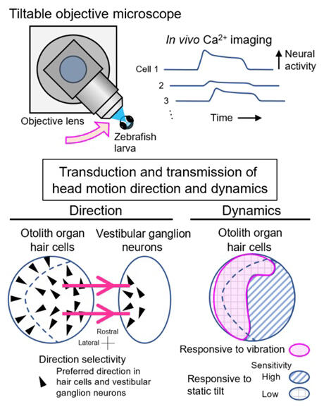
The researchers used their tilting objective microscope to visualize the hair cells of the utricle, which is thought to be sensitive to horizontal plane movements and tilting in the anterior-posterior and lateral directions in the otolith organ of zebrafish larvae.
Specifically, the group created genetically modified fish expressing the green fluorescent Ca2+ sensor (GCaMP) and red fluorescent protein in the hair cells, and conducted calcium imaging of all hair cell activity while the fish were tilted back and forth and side to side. When tilted, they observed an increase in the fluorescence intensity ratio in hair cells at different locations depending on the tilting direction, and visualized direction-preferential activity. When the directional preference of individual hair cells was expressed as a vector, the direction was consistent with hair-bundle polarity. Furthermore, they found that the magnitude of the response varied from place to place within the otolith organ.
Provided by NIBB
"Using this microscope, we will be able to visualize neural activity in the brain just as well as in the inner ear, and we are looking forward to further research into how sensory information about head movement and gravity is processed in neural circuits, and how posture is controlled," explained Tanimoto.
Journal Information
Publication: Nature Communications
Title: Tiltable objective microscope visualizes selectivity for head motion direction and dynamics in zebrafish vestibular system
DOI: 10.1038/s41467-022-35190-9
This article has been translated by JST with permission from The Science News Ltd. (https://sci-news.co.jp/). Unauthorized reproduction of the article and photographs is prohibited.