A research group led by Professor Kazunari Ishii of the Department of Medicine, Kindai University, used a prototype of the BresTome TOF-PET system — the world's first high-resolution PET system specialized for head and breast examinations, developed by Shimadzu Corporation — to produce higher quality (higher resolution) images than conventional systems, demonstrating the system's usefulness in diagnosis. The group hopes that the device's higher-quality images will contribute to the early treatment of dementia, which requires accurate diagnosis. A paper on the group's findings was published on January 4 in the Journal of Nuclear Medicine.
Unlike CT or MRI scans, which examine morphological abnormalities, PET scans are functional imaging tests that examine metabolism and other functions. For example, PET scans can image glucose metabolism and the accumulation of substances in the brain that cause Alzheimer's disease.

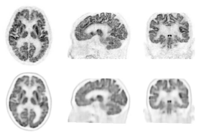
Bottom: Brain FDG-PET Images Obtained with Conventional System
Top images appear of higher resolution and show greater detail. Bottom images appear somewhat blurry.
Provided by Shimadzu Corporation
However, functional imaging systems such as PET have poor resolution and blurred images compared to morphological imaging. For this reason, a PET system capable of capturing high-resolution images has been highly sought-after.
The Faculty of Medicine at Kindai University and Shimadzu have been conducting clinical research at Kindai University Hospital since October 2020 using a prototype of the company's BresTome TOF-PET system. It is the world's first new high-resolution PET system that can image the breast and the head.
In the clinical study, patients with dementia were tested mainly for glucose metabolism and amyloid deposition. The team compared images taken with the high-resolution PET system with those taken with a conventional PET/CT system. Results showed that in all cases, the resolution of the images taken with the high-resolution PET system was superior.
In some cases, it also led to a change in diagnosis. In about 10% of cases in which brain amyloid PET scans are used, results are difficult to determine conclusively because of ambiguous accumulations in gray matter. In the present study, amyloid deposition was confirmed in the right temporal lobe by conventional PET/CT equipment in one out of 17 patients.
This result would be a positive diagnosis for amyloid deposition (Alzheimer's pathology present) using radiogram interpretation guidelines. However, imaging these patients with the high-resolution device indicated no amyloid deposition in any area, including the right temporal lobe. This meant the result was negative and thus changed their diagnoses.
Thus, even in clinical use, this high-resolution PET system was shown to enable accurate diagnoses and contribute to the early treatment of dementia.
This article has been translated by JST with permission from The Science News Ltd. (https://sci-news.co.jp/). Unauthorized reproduction of the article and photographs is prohibited.


