
A team of researchers led by Postdoctoral Researcher Rachana Ouk and including Assistant Professor Takao Oi, Lecturer Daisuke Sugiura, and Professor Mitsutaka Taniguchi of the Graduate School of Bioagricultural Sciences, Nagoya University, has used the serial sectioning method to create a three-dimensional reconstruction of rice leaves. The method involves cutting samples at regular intervals for observation. By analyzing the 3D data, the team could accurately measure the surface area of mesophyll cells and chloroplasts facing intercellular air spaces, which are closely related to the photosynthetic capacity of the plant.
Provided by Nagoya University
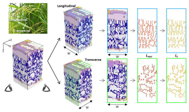
A critical component of photosynthesis, the exchange of carbon dioxide, occurs through the surface areas of cells and chloroplasts facing the intercellular spaces. These areas have traditionally been determined by analyzing planar images of thinly sliced leaf tissue under a microscope. According to Oi, "In rice leaf tissue, cross-sectional slices show curved cell sections at the interface, and longitudinal slices show cell sections aligned in a palisading pattern, so there was no clear indicator from which to estimate the surface area."
When estimating photosynthetic gas exchange capacity from leaf structure, a crucial parameter is the mesophyll and chloroplast surface area per unit of leaf area, which represents the area of chloroplasts facing either the mesophyll cell walls or intercellular space. The research group obtained a three-dimensional image of rice leaf tissue by cutting the tissue into 100 to 200 sections at 0.5-micrometer intervals and stacking planar images taken under a microscope. They then measured the volume of cells and chloroplasts and the surface area facing the intercellular space. The 3D data was then used to create horizontal and vertical cross-sections. Results demonstrate the conditions under which the difference between the measured values and those estimated using the conventional method are more minor.
After comparing the 3D-measured mesophyll surface areas and chloroplast surface areas with the 2D estimated values, the group found that the estimate was significantly impacted by the direction of the cut and curvature correction factor, leading to a deviation of 10 to 40% from the 3D measured values. Therefore, when estimating chloroplast and mesophyll surface area from 2D cut sections of rice leaf tissue, the average value most closely approximates the 3D measured value when assuming the mesophyll cells to be oblate using longitudinal slices.
The surface area of chloroplasts and mesophyll cells remained the same regardless of the calculation method, with mesophyll cells accounting for approximately 90% of the surface area of the chloroplast cell wall in contact with the intercellular space. Chloroplasts in rice leaves are thought to enhance gas exchange capability by being arranged along the intricately curved cell walls, covering a significant portion of the surfaces that face the intercellular spaces within the cells.
Oi expects that the results of the recent study will enhance the accuracy of rapid estimation methods and contribute to research aimed at increasing photosynthetic capacity in crops.
Journal Information
Publication: Annals of Botany
Title: 3-D reconstruction of rice leaf tissue for proper estimation of surface area of mesophyll cells and chloroplasts facing intercellular airspaces from 2-D section images
DOI: 10.1093/aob/mcac133
This article has been translated by JST with permission from The Science News Ltd. (https://sci-news.co.jp/). Unauthorized reproduction of the article and photographs is prohibited.