
A research group led by Research Associate Toshiyuki Takagi and Professor Koji Inoue of the Atmosphere and Ocean Research Institute, the University of Tokyo, Postdoctoral Researcher Keisuke Motone of the University of Washington, Professor Hideyuki Yamashiro of the University of the Ryukyus, and Assistant Professor Natsuko Miura of the Graduate School of Agriculture, Osaka Metropolitan University, has discovered a photoprotective pigment bacterium on the cell surface of dinoflagellates that live in corals and shellfish, and that increasing the amount of these bacteria improved the resistance of dinoflagellates to intense light stress. Increasing the abundance ratio and the number of viable dinoflagellates pigment bacteria ensured that high photosynthetic capacity could be maintained even under high light stress conditions. The findings are anticipated to lead to the protection and conservation of corals and dinoflagellates using probiotics. The group's research was published in the January 18 issue of Microbiology Spectrum.
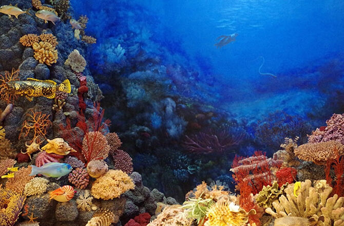
Humans benefit from ecosystem services found on coral reefs, which are home to about 25% of all marine species and form a rich biodiversity comparable to that on land.
In recent years, however, rising seawater temperatures and intense light stress have led to a bleaching phenomenon in which the dinoflagellates symbiotic with corals disappear. Bacteria co-existing on corals have been reported to have the ability to inhibit this bleaching, but the function and localization of these bacteria has remained a mystery.
So far, the research group has discovered a pigment bacterium that synthesizes carotenoids that co-occur with dinoflagellates and alleviate high temperature and intense light stress. When various bacteria co-existing in the process of isolating and culturing dinoflagellates from corals were removed with antibiotics, these pigmented bacteria were found to survive. The research group therefore sought to identify the interactions between dinoflagellates and co-existing pigment bacteria.
First, they treated dinoflagellates isolated and cultured from corals and shellfish for 50 days in a medium containing the antibiotics kanamycin, ampicillin and streptomycin. After recovering and culturing them in an antibiotic-free medium again, the researchers conducted an analysis of 16S rRNA gene amplicon sequence to compare the bacterial flora (types of bacteria constituting the flora) before and after antibiotic treatment.
This revealed a trend towards an increase in pigmented bacteria in all dinoflagellates, confirming the tendency for only pigmented bacteria to survive in the dinoflagellates flora after antibiotic treatment. In particular, 100% of the shell derived Cladocopium dinoflagellate strains were Maribacter bacteria that synthesize orange pigments. In addition, 90% of the Durusdinium dinoflagellate strains derived from thistle coral were Roseivirga bacteria that synthesize pink pigments.
Having successfully isolated the pigmented bacteria, these two types of pigmented bacteria were cultured in media containing each of the three antibiotics alone. Initially, each pigment cell was expected to be resistant to all antibiotics based on the process of previous manipulations, but both bacteria were sensitive to ampicillin. The researchers thought that ampicillin resistance occurred in co-existence with dinoflagellates.
When they examined the distribution of pigment bacteria in dinoflagellates to investigate the cause of this, all pigment bacteria were localized on the surface of the dinoflagellates. It indicated that the bacteria may have escaped the action of the antibiotics. Further examination of Maribacter bacteria, which had higher pigment levels, revealed that they synthesize zeaxanthin (a type of carotenoid pigment), which has photoprotective and antioxidant functions.
These results suggest that the co-existence of pigmented bacteria on the cell surface of dinoflagellates may reduce light stress. The group observed the photosynthetic capacity (Fv/Fm) of the dinoflagellates, which had been manipulated to 100% of Maribacter genus bacteria and eight times the number of viable bacteria, continuously for 14 days under strong light stress conditions and compared them with normal dinoflagellates and no stress conditions.
They found that dinoflagellates engineered with Maribacter under high light stress conditions maintained a higher photosynthetic capacity than normal dinoflagellates. On the other hand, normal dinoflagellates maintained higher photosynthetic capacity than Maribacter bacteria manipulated dinoflagellates under stress-free conditions, indicating that other bacteria may contribute to maintaining photosynthetic capacity.
"Moving forward, we would like to investigate whether the photoprotective function of the pigment bacteria actually functions in living corals rather than in culture, and whether it leads to suppressing coral bleaching," commented Takagi.
Journal Information
Publication: Microbiology Spectrum
Title: Mutualistic interactions between dinoflagellates and pigmented bacteria mitigate environmental stress
DOI: 10.1128/spectrum.02464-22
This article has been translated by JST with permission from The Science News Ltd. (https://sci-news.co.jp/). Unauthorized reproduction of the article and photographs is prohibited.