
A research group led by Assistant Professor Eisuke Kanao, Professor Yasushi Ishihama and Project Leader Jun Adachi of the Practical Drug Discovery Research Project between the Graduate School of Pharmaceutical Sciences of Kyoto University and the National Institutes of Biomedical Innovation, Health and Nutrition (NIBIOHN), a group led by Associate Professor Takuya Kubo, Professor Koji Otsuka and graduate student (at the time) Shuntaro Wada of the Graduate School of Engineering at Kyoto University, and a group led by Professor Kazunari Akiyoshi, have developed a sponge‐like polymer separation medium that can classify extracellular vesicles, microparticles released from cells, by recognizing the sugar chains attached to their surfaces, and have successfully revealed that even extracellular vesicles derived from the same cell function differently depending on the sugar chains on their surface.
Provided by Kyoto University
Lectin‐based affinity chromatography (LAC) using glycan‐recognition proteins (lectins in research) as stationary phases is a typical separation method for glycans, but conventional separation materials for LAC have small pores, and microparticles such as extracellular vesicles are larger than these pores, so they clog the base material and cannot pass through.
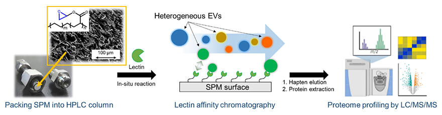
The group has independently developed a sponge‐like polymer separation base material (sponge monolith) with pores 10‐100 µm larger than those of conventional base materials to achieve high water permeability and used it as a separation base material for LAC, thereby solving this problem.
Specifically, using sialic acid‐recognising Sambucus sieboldiana agglutinin (SSA) and mannose‐recognising concanavalin A (ConA) as models, spongy monoliths were prepared with each lectin immobilized, and attempts were made to separate extracellular vesicles recovered from cultured cells (HEK293 cells) by ultracentrifugation into subclasses.
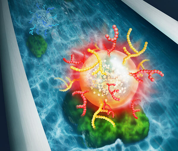
Extracellular vesicles with specific surface glycans corresponding to each lectin were successfully recovered intact, without loss of shape. Furthermore, proteome analysis revealed that the constituent proteins and their functions differed significantly between extracellular vesicles recovered with SSA and those recovered with ConA.
Copyright 2023 American Chemical Society.
"Conventional separation materials have small through‐pores and are not suitable for separating large structures such as extracellular vesicles, but we have overcome this problem by applying a proprietary porous sponge polymer as a separation material," explained Kanao. "This work has the potential to become a fundamental technology for extracellular vesicle separation in drug discovery research, and we expect the application of this concept to create a platform for the separation of all biologically relevant particles, including viruses."
Journal Information
Publication: Analytical Chemistry
Title: Classification of Extracellular Vesicles Based on Surface Glycan Structures by Spongy‐like Separation Media
DOI: 10.1021/acs.analchem.2c04391
This article has been translated by JST with permission from The Science News Ltd. (https://sci-news.co.jp/). Unauthorized reproduction of the article and photographs is prohibited.