
Neurons in the cerebral cortex are known to be transformed by various experiences, but the deepest location, layer 6b, has been difficult to measure and it was a mystery as to whether neurons in this layer undergo transformation.
Project Assistant Professor Taisuke Yoneda and Professor Yumiko Yoshimura from the National Institute for Physiological Sciences and their colleagues succeeded in measuring neurons in layer 6b of the cortex's primary visual cortex in mice and found that their functional features are altered by visual experiences after birth. Their research was published in PNAS.
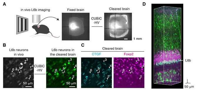
(B) L6b neurons from in vivo two‐photon imaging (left) and the same areas from a cleared brain (right).
(C) Most of the recorded L6b neurons expressed CTGF which is a subplate neuron marker. Foxp2 is a marker of cortico‐thalamic neurons.
(D) An example of a volumetric image of cleared brain.
Provided by the National Institute for Physiological Sciences
Neuronal cells change dramatically in the cortex before and after birth, and subplate neurons play an important role in the formation of the neural network. Many subplate neurons are lost by cell death towards the end of the formation of the network, but some of them are known to survive in layer 6b, the deepest part of the cortex. However, as layer 6b is deep in the cortex, it is difficult to measure them and the function of surviving subplate neurons has been a mystery. By optimizing the reagents used in their experiments, the research group succeeded in making two‐photon calcium imaging measurements from layer 6b.
Furthermore, using tissue transparency and antibody staining, they found that more than 70% of the neurons in layer 6b where calcium imaging measurements were made expressed the subplate neuron marker protein.
The researchers subsequently investigated the functional properties of subplate neurons and found that they, like other primary visual cortex neurons, have functional features such as orientation selectivity and spatial frequency selectivity. They then investigated whether the neurons in layer 6b change with experience.
The group used two‐photon imaging to investigate whether ocular dominance plasticity induced by changes in visual experience during development also occurs in layer 6b. After blocking input from one eye in 26‐day‐old mice, they found that three days later, fewer cells responded to input from the blocked eye. These findings indicate that layer 6b contains neurons that undergo experience‐dependent changes, resulting in ocular dominance plasticity.
"In this study, we were able to shed light on some of the functions of layer 6b neurons in areas induced by changes in ocular experience during visual development." commented Yoneda. "Going forward, it will be important to examine the function of 6b neurons in the auditory and somatosensory cortex to determine whether there are similarities and differences in function. Subplate neurons that remain in the brain after birth may flexibly reconfigure their neural circuits in response to the postnatal environment and co‐ordinate with other neurons to optimize information processing in the cortex."
Journal Information
Publication: Proceedings of the National Academy of Sciences of the United States of America (PNAS)
Title: Experience‐dependent functional plasticity and visual response selectivity of surviving subplate neurons in the mouse visual cortex
DOI: 10.1073/pnas.2217011120
This article has been translated by JST with permission from The Science News Ltd. (https://sci-news.co.jp/). Unauthorized reproduction of the article and photographs is prohibited.