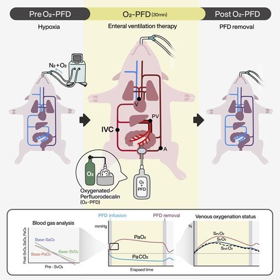
Respiratory failure' is a serious condition where one's own respiratory function declines due to various diseases such as COVID‐19. During respiratory failure, oxygen and carbon dioxide concentrations decrease and increase, respectively, in the blood beyond the normal ranges. Using a porcine model, a joint research group comprising of Tokyo Medical and Dental University (TMDU) and Nagoya University demonstrated the effectiveness of the 'enteral ventilation (commonly known as EVA)'; the method is expected to be a completely new adjunct therapy for respiratory disorders. The research was supported by the Japan Agency for Medical Research and Development (AMED), Grants‐in‐Aid for Scientific Research (Kakenhi) and crowdfunding donations and was published in the international scientific journal iScience.

Provided by TMDU
The achievement was brought about by the joint research group comprising of Professor Takanori Takebe of the Institute of Research at Tokyo Medical and Dental University, Lecturer Tasuku Fujii of the Department of Anesthesiology at Nagoya University Hospital, Professor Kimitoshi Nishiwaki of the Department of Anesthesiology at Nagoya University Graduate School of Medicine and Professor Toyofumi Chen‐Yoshikawa at the Department of Thoracic Surgery at Nagoya University Graduate School of Medicine.
Respiratory care by ventilators or care by extracorporeal membrane oxygenation (ECMO) are used in patients with severe 'respiratory failure'; however, sometimes these can cause lung disorders, bleeding/thrombosis in vital organs and systemic infections. Such severe complications remain as issues. In addition, these methods require advanced medical equipment and human resources, and the collapse in the supply‐demand balance during a pandemic restricts access to medical resources.
The COVID‐19 situation has highlighted that many lives were lost under these circumstances. Therefore, at the forefront of medicine, there has been a strong demand for novel respiratory support therapies that are safer, less invasive, and allow for the easier achievement of oxygenation and ventilation effects.
To solve these problems, the Takebe group focused on the fact that aquatic organisms, such as loaches, perform respiration not only using the gills and skin but also through the intestine to support respiratory function. Previous studies have reported that 'enteral ventilation' — that is, respiration via the intestinal tract — is possible even in mammals such as mice. This time, collaborating with the team of Fujii, Nishiwaki and Yoshikawa (experts in anesthesia and respiratory management) and others, they used a pig as a large animal model with respiratory failure to reproduce conditions similar to humans and to examine the effectiveness of the 'enteral ventilation.'
First, they established a hypoxic model of respiratory failure in pigs under anesthesia and muscle relaxants and controlled its respiratory function via artificial respiration. They then selected rectal catheters and perfluorodecalin (PFD), which have been used in clinical settings, and developed an administration protocol that has clinical applications. Subsequently, the PFD was oxygen‐bubbled in advance and was administered through a catheter inserted into the anus, as in an enema. The oxygen concentration and carbon dioxide partial pressure in the blood were monitored to determine the ventilation effect through the intestinal tract.
As a result, it was discovered that the administration of oxygenated PFD into the intestinal tract not only improved hypoxia but also reduced carbon dioxide levels, indicating that the ventilation effect had been achieved. Additionally, it was proven that the effect was improved by the administration of PFD as the original state returned after its excretion. Furthermore, in order to identify the blood vessel responsible for oxygenation, catheters were placed in multiple venous circulations, and the oxygenation status was evaluated. Multiple blood vessels from the intestinal tract were found to be oxygenated.
During these processes, no serious side effects were observed in hemodynamics (blood pressure and pulse) or the state of the intestinal tract and spleen. As such, it was demonstrated that the 'enteral ventilation' could be performed safely. In the future, the joint research group will aim for the clinical application of the new 'enteral ventilation' and confirm its efficacy and safety in humans as a medical device. Clinical research is already in progress with EVA Therapeutics (a venture from Tokyo Medical and Dental University) and Maruishi Pharmaceutical.
Journal Information
Publication: iScience
Title: Enteral liquid ventilation oxygenates a hypoxic pig model
DOI: 10.1016/j.isci.2023.106142
This article has been translated by JST with permission from The Science News Ltd. (https://sci-news.co.jp/). Unauthorized reproduction of the article and photographs is prohibited.


