
The joint research group led by Professor Hideyuki Okano, Team Leader of the Laboratory for Marmoset Neural Architecture at the RIKEN Center for Brain Science (also affiliated with the Department of Physiology, Keio University Graduate School of Medicine), along with Visiting Scientist Junichi Hata (Associate Professor, Graduate School of Human Health Sciences, Tokyo Metropolitan University), and Project Associate Professor Ken Nakae of ExCELLS at the National Institutes of Natural Sciences have developed and released a digital brain database for common marmosets (small primates) using magnetic resonance imaging (MRI). The Innovative Brain Project aims to contribute to the elucidation of the macro‐level neural circuits responsible for the higher brain functions of primates, overcoming psychiatric and neurological disorders in humans, and advancing information processing technology. The results were published in the online version of Scientific Data.
Provided by RIKEN
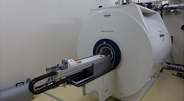
The joint research group first established methods for stably performing MRI experiments on common marmosets under anesthesia, collecting multi‐contrast MRI data, and processing the images, and subsequently collected MRI data of the brains of common marmosets and evaluated their characteristics. The study included a total of 216 healthy common marmosets (88 males and 128 females) with an average weight of 357.1 ± 60.2 g. The age of the marmosets ranged from 0.8 to 10.3 years, with an average age of 4.34 ± 2.56 years. The MRI apparatus was equipped with an ultrahigh‐field magnet of 9.4 T and a transmitting and receiving coil with an inner diameter of 86 mm.
The group conducted three measurements to obtain multi‐contrast MRI information of the common marmoset brain. The first measurement involved acquiring T1‐weighted and T2‐weighted anatomical images, and the optimized magnetic field and radio wave pulse programs were required to align with the 9.4 T MRI equipment. The second measurement involved acquiring diffusion‐weighted images with network information on nerve fiber connections, and the third measurement involved observing resting brain activity to obtain network information on brain activity.
Among the obtained MRI data, the information on the nerve fiber network and brain activity could not be utilized directly; it required image processing and analysis to extract network information. These image analysis methods were also optimized for the common marmoset brain.
The MRI data and analysis results obtained were uploaded to the website, providing an open‐access digital brain MRI data. The database is classified according to various characteristics, such as the age, gender, and physique (weight) of common marmosets. On the database site, users have the capability to extract and download MRI digital brain data by using filters based on relevant information such as age, gender, and weight. In addition, a preview function is provided, and brain images can be checked for each marmoset. The brain is divided into various areas, including the motor area, sensory area, auditory area, thalamus, striatum, and more. Territory atlases for these areas are provided so that brain scientists can download and utilize the results.
The multi‐contrast MRI database of common marmosets provides not only highly reliable and detailed brain structural and functional information but also essential knowledge about the effects of brain aging. The MRI data of marmosets is valuable for understanding the comprehensive macro‐level neural circuits, including higher brain functions, in primates. They also offer crucial insights into the effects of brain aging when combined with disease models and human MRI data.
At the time of publication, this database represented the world's largest public database of common marmoset brain data. It serves as a valuable resource for understanding the effects of various factors, such as age, gender, and body type, on the brain.
This article has been translated by JST with permission from The Science News Ltd. (https://sci-news.co.jp/). Unauthorized reproduction of the article and photographs is prohibited.