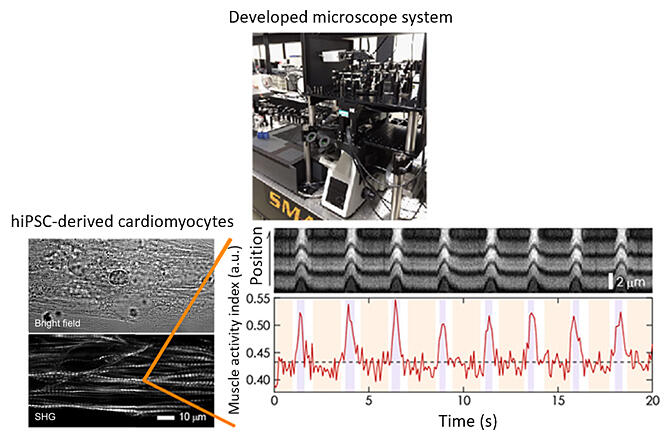
Currently, few technologies can directly evaluate muscle activity in live cells and tissues. A joint research group led by Team Leader Tomonobu Watanabe of the Laboratory for Comprehensive Bioimaging, RIKEN Center for Biosystems Dynamics Research (Professor at Hiroshima University), Assistant Professor Hideaki Fujita of the Research Institute for Radiation Biology and Medicine of Hiroshima University, Professor Shigeru Miyagawa of the Osaka University Graduate School of Medicine, and Professor Erina Kuranaga of the Tohoku University Graduate School of Life Sciences has developed a technology for the non-contact, non-invasive quantitative evaluation of muscle activity in live cells and tissues. The technology will contribute to the quality control of artificial cardiomyocytes derived from induced pluripotent stem (iPS) cells, diagnosing heart disease, and studying individual differences in the effects of radiation exposure. The research has been published in the online edition of Life Science Alliance.

Provided by RIKEN
The joint research group has developed a measurement and analytical method that uses optical second-harmonic generation (SHG), a nonlinear optical phenomenon, to be able to estimate the activity of myosin, a protein that acts when muscle fibers contract inside cells or tissues.
SHG light is generated by the reflection of the permanent electric dipole moments and their arrangement in the illuminated object. In other words, the observed SHG light changes when the protein structure changes. Studies have shown that the polarization characteristics of SHG light from myofibrils (SHG anisotropy) differ between the rigid and relaxed states of muscle. However, no reports were available on measuring SHG anisotropy within a single myocardial cell and during cardiomyocyte beating. This is due to a lack of measurement sensitivity.
In 2019, the RIKEN team developed the world's most sensitive SHG polarized light microscope. Using this microscope, the team observed cardiomyocytes differentiated from healthy human-derived iPS cells and was able to visualize muscle structures while keeping the cells alive selectively. Given that 12.5 images can be acquired per second (temporal resolution: 80 ms), the SHG polarization of cardiomyocytes can be accurately measured even when they beat several times per second.
The collaborative research group has succeeded for the first time in directly evaluating pulsed myosin activity in synchrony with a myocardial beating by calculating an index (γ value; representing myosin activity) from the polarization characteristics of the acquired SHG light.
Using this method, they were also able to assess myocardial dysfunction quantitatively in iPS cell-derived cardiomyocytes from patients with heart disease and the effect of genome editing on repair (therapy) and late-onset (occurring a certain time after exposure) cardiac dysfunction in iPS cell-derived cardiomyocytes after UV irradiation.
Additionally, Drosophila models of Barth syndrome (caused by mutations in the tafazzin-encoding gene) were used to challenge the assessment of muscle dysfunction because SHG measurements use near-infrared light (which has high biological transparency) as the incident light.
Using Drosophila pupae, the researchers could non-invasively assess myosin activity in the internal body wall muscles. Myosin activity was reduced in pupae from the Barth syndrome model. This is the first experimental example of the direct assessment of muscle activity inside a live Drosophila body.
The measurement and analytical method is expected to become an indispensable research tool for studying the mechanobiology of cardiomyopathies, iPS cells, and the effects of late-onset radiation exposure.
Journal Information
Publication: Life Science Alliance
Title: Estimation of crossbridge-state during cardiomyocyte beating using second harmonic generation
DOI: 10.26508/lsa.202302070
This article has been translated by JST with permission from The Science News Ltd. (https://sci-news.co.jp/). Unauthorized reproduction of the article and photographs is prohibited.


