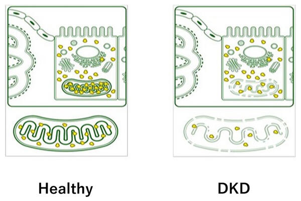
Associate Professor Kazuhiro Hasegawa and Professor Shu Wakino of the Graduate School of Biomedical Sciences at Tokushima University and their research group discovered that activation of mitochondrial ribosomes activates the mitochondria, reduces urine protein levels in a mouse model with diabetic kidney disease and improves kidney disorder, all of which are new therapeutic implications. Their achievements were published in the Journal of the American Society of Nephrology (JASN).

Provided by Tokushima University
Diabetes mellitus is a common lifestyle disease, affecting an estimated 10 million patients in Japan. Diabetic nephropathy or diabetic kidney disease is the leading cause for introducing dialysis. Treatment of diabetic kidney disease mainly involves treatment for diabetes and hypertension, and there is no effective treatment for the kidney itself. This has meant that there is no way to halt the increasing number of patients and medical care bills related to this disease.
Kidneys produce urine from the inflowing blood and excrete waste products from the body. The glomeruli, kidney structures that filter the blood and produce urine, have a yarn ball-like structure consisting of a mass of capillaries. If the filter becomes congested, urine is not produced, and if the protein component is not filtered out proteinuria arises. Kidneys have kidney tubules where filtered urine (primary urine) passes through. In this area, necessary substances are reabsorbed from the primary urine, and waste products remain in the primary urine for excretion. Through this, urine is produced and eventually excreted from the body. Mitochondria and mitochondrial ribosomes are abundantly distributed in the kidney tubules.
In a previous study, the research team found that, in the kidney, metabolic imbalance in the kidney tubules, which was previously regarded as the urinary passageway, is also associated with the abnormal function of cells called podocytes (which make up the filter of the glomerulus). They further clarified the order of disease progress, including damage to the filter and the appearance of proteinuria. Loss of communication from kidney tubular cells to glomerular podocytes at a very early stage of diabetes results in the onset of the disease.
The research team newly revealed that mitochondrial ribosome hypofunction is involved in energy metabolism imbalance of kidney tubules. When mitochondrial ribosome hypofunction occurs, diabetic kidney disease has already developed. They also discovered the possibility that a new treatment that replenishes the enzyme phosphoenolpyruvate carboxykinase 1 (PCK1), where primary focus has been on its action as a gluconeogenic enzyme, may be effective in repairing the failure.
More specifically, when the PCK1 gene was overexpressed only in kidney tubules of mice with diabetic nephropathy, a proteinuria-reducing effect, which has been thought to occur mainly due to the failure of filtering function of glomeruli, was observed, and they discovered that the activation of mitochondria ribosomes was a key factor in the underlying mechanism. In other words, they found that the development of a drug that activates PCK1 held the clue to an epoch-making therapy that can suppress the progression of diabetic kidney disease in the long term.
Once diabetic kidney disease progresses to a certain extent, it is difficult to stop further progression. It is expected that a high degree of efficacy can be obtained by implementing pre-emptive medicine that controls the onset of the disease and also avoids the intensification of the severity, rather than conventional treatments that delay the onset and progression. Advances in this research are expected to lead to the development of new treatments for humans through ultra-early intervention.
Journal Information
Publication: Journal of the American Society of Nephrology
Title: PCK1 Protects against Mitoribosomal Defects in Diabetic Nephropathy in Mouse Models
DOI: 10.1681/ASN.0000000000000156
This article has been translated by JST with permission from The Science News Ltd. (https://sci-news.co.jp/). Unauthorized reproduction of the article and photographs is prohibited.


