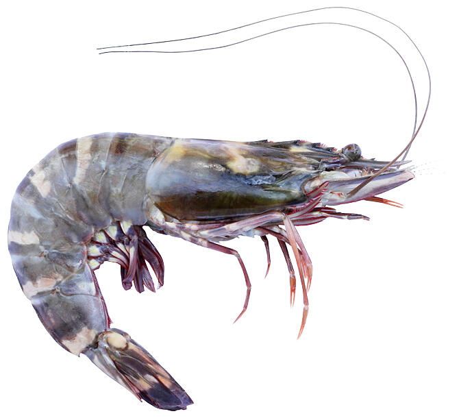
Viral diseases have become a problem at sites where tiger prawn, white-legged prawn (white-leg shrimp), bull prawn (black-tiger shrimps), and other types of prawn that are often seen on dining tables are farmed. As prawns do not have adaptive immune systems (i.e, the ability to produce antibodies), vaccines are ineffective against viral infections in these invertebrates, unlike in vertebrate mammals and fish. There are currently no effective means of determining which cells of the prawn are affected by a virus and why they can no longer fight the pathogen, because the blood cells controlling the immunity of prawns cannot be objectively classified through microscopic observation.
The research group led by Assistant Professor Keiichiro Koiwai of the Department of Marine Bioresources at Faculty of Marine Science and Technology at Tokyo University of Marine Science and Technology has succeeded in identifying the cell populations and genes that are affected by viral infections. They achieved this using comprehensive single-cell mRNA analysis with a microfluidic device, and they published these results in the online edition of Marine Biotechnology.

The research group artificially infected tiger prawns with white spot syndrome virus, which causes severe damage to prawn farming. Then, they screened severely infected prawn individuals using real-time PCR, where blood cells were encapsulated within microdroplets in a microfluidic device, and comprehensive single-cell mRNA analyses were performed. Gene expression data for 3724 cells derived from healthy individuals and 3064 cells derived from virus-infected individuals were collated and compared using bioinformatics analysis. Consequently, it was clarified that the ratio of the antimicrobial peptide-producing cell population decreased to approximately 15% as a result of the virus infection, whereas it accounted for approximately 25% in the healthy state. The researchers further identified multiple marker genes that enable the objective classification of blood cells of tiger prawns, regardless of the presence or absence of virus infection and succeeded in microscopically observing the mRNA expression patterns in the prawn individuals.
Although fresh cells are usually required for single-cell mRNA analysis, by applying the methanol preservation method (effective for human leukocytes) to prawn hemocytes, it was additionally possible to perform sampling on a different day. In this way, the researchers achieved comprehensive single-cell mRNA analyses and confirmed that the data were comparable to those of live cells. This allows both sampling and experimental manipulation to be performed remotely.
Now that the blood cell population that decreases during a virus infection has been found, it is expected that further research will lead to the conquering of viral diseases and improvements in prawn breeding methods and feed additives. Additionally, by conducting exhaustive single-cell mRNA analyses, a picture of the cells that make up an organism's body (cell Atlas) can be constructed—even those of cultured fish and shellfish, whose cell classification remains problematic compared with that of model organisms such as tiger prawns. Such an Atlas may form the reference basis for developing new countermeasures against fish diseases.
This article has been translated by JST with permission from The Science News Ltd. (https://sci-news.co.jp/). Unauthorized reproduction of the article and photographs is prohibited.


