
X-ray CT can be used to visualize the internal features of an object in 3D by capturing projected images of the object from multiple directions. By capturing more than 30 frames of an object in continuous shots per second, corresponding videos can be created. The X-ray CT equipment used in hospital settings requires several seconds to several tens of seconds to capture images of a sample. This imaging speed can be increased using powerful X-ray beams of synchrotron radiation. However, to capture the projected images of a sample from various directions, the sample must be rapidly rotated.
For example, to perform X-ray CT with a time resolution of 1 millisecond, the sample must be rotated at a high speed of 30,000 rotations per minute. Because such rapid sample rotations may cause sample deformation owing to centrifugal forces and may not be suitable for fluid samples, several challenges are encountered in controlling sample environments. Consequently, even when using synchrotron radiation, the time resolution of conventional 4D-X-ray CT is approximately 10 milliseconds.
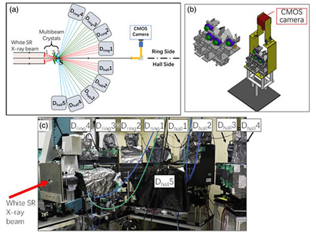
A research team led by Professor Wataru Yashiro of the International Center for Synchrotron Radiation Innovation Smart (SRIS) at Tohoku University succeeded in the world's first proof-of-principle demonstration of 4D-X-ray CT with a temporal resolution of 0.5 milliseconds (a spatial resolution of approximately 10 micrometers), which exceeds 1 millisecond. This was achieved by developing innovative optical elements to split the multiplex synchrotron radiation into approximately 30 beams, a multi-beam image detector to simultaneously capture all projected images, and an advanced compressed-sensing-based CT reconstruction algorithm to enable 3D visualization with fewer projections. The research results have been published in Applied Physics Express.
Provided by Tohoku University
This technology has enabled the acquisition of 4D-X-ray CT images of phenomena that were otherwise impossible to capture using conventional methods. Thus, the potential impact of this development extends to various fields, from academic research to industrial applications, including material destruction, fluid and viscoelastic material behavior, machining, abrasion, welding and combustion.
Journal Information
Publication: Applied Physics Express
Title: Sub-millisecond 4D X-ray tomography achieved with a multibeam X-ray imaging system
DOI: 10.35848/1882-0786/ace0f2
This article has been translated by JST with permission from The Science News Ltd. (https://sci-news.co.jp/). Unauthorized reproduction of the article and photographs is prohibited.