
Assistant Professor Yusuke Nasu, Technician Yuki Kamijo, and Professor Robert E. Campbell of the Graduate School of Science at the University of Tokyo, and their colleagues, have developed a biosensor that observes lactate in living mice by making full use of protein engineering techniques. The series of lactate biosensors, named LACCO, has the highest known sensitivity as a genetically encoded sensor, captures the dynamics of lactate inside and outside cells, and is a powerful tool for revealing a new role for lactate. These developments have been published in Nature Communications.
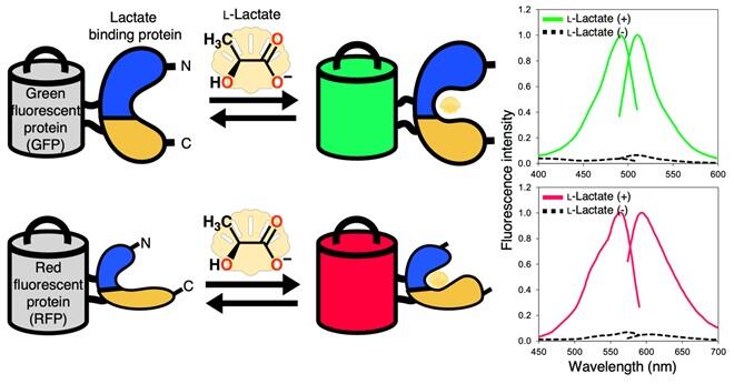
A green fluorescent extracellular L-lactate biosensor eLACCO2.1 (upper) and a red fluorescent intracellular L-lactate biosensor R-iLACCO1 (bottom). Excitation and emission spectra of eLACCO2.1 and R-iLACCO1 in the presence and absence of L-lactate are shown in the right graphs.
Provided by the University of Tokyo
Although the human brain accounts for only approximately 2% of the total body weight, it consumes approximately 20% of the total energy ingested. For a long time, it has been believed that glucose is the main energy substance for nerve cells, and that lactate is a simple metabolic byproduct. However, recent theory suggests that lactate is used as an energy substance in nerve cells. Therefore, the role of lactate needs to be reconsidered, and observations of lactate dynamics inside and outside cells are required to verify this theory.
The research group had previously developed an extracellular lactate biosensor (eLACCO1.1) (Nasu et al. Nat. Commun. 12, 7058, 2021); however, the sensitivity was insufficient for its application to living animals. In addition, visualizing intracellular lactate dynamics in living organisms was still challenging with the original biosensor.
The research group developed an extracellular lactate sensor (eLACCO2.1) and an intracellular lactate sensor (R-iLACCO1) using a protein engineering technique called directed evolution. To visualize extracellular lactate, a sensor molecule was devised to be expressed on the cell membrane surface. This lactate sensor has the highest known sensitivity, 100 times more sensitive than that of conventional sensors in terms of fluorescence signal changes in response to changes in lactate concentrations, and the research group has successfully visualized the lactate dynamics inside and outside neurons in the brain of living mice. By simultaneously using different fluorescence wavelengths (green outside the cell, red inside the cell), they also succeeded in concurrently visualizing the lactate dynamics inside and outside the cell.
Their experiments successfully demonstrated how insulin stimulation of live mice reduces lactate concentration outside nerve cells and how stimulation of whiskers with air changes the lactate concentration inside nerve cells. Lactate has long been viewed negatively as a metabolic byproduct and muscle fatigue agent. Research using the LACCO series is expected to demonstrate that lactate is an extremely important energy source for brain activity.
Journal Information
Publication: Nature Communications
Title: Lactate biosensors for spectrally and spatially multiplexed fluorescence imaging
DOI: 10.1038/s41467-023-42230-5
This article has been translated by JST with permission from The Science News Ltd. (https://sci-news.co.jp/). Unauthorized reproduction of the article and photographs is prohibited.