The combined research group of Director Kazuhiko Seki and Section Chief Shinji Kubota of the Department of Neurophysiology at the National Institute of Neuroscience (NIN) of the National Center of Neurology and Psychiatry (NCNP), and Project Associate Professor Saeka Tomatsu of the Division of Behavioral Development at the National Institute for Physiological Sciences (NIPS) has revealed that the sensory signals generated in the limbs during exercise are regulated by a brain mechanism called "presynaptic inhibition," which allows for skillful control of movement. The researchers analyzed the spinal cord of a monkey performing wrist flexion and extension and found that by manipulating "presynaptic inhibition," the brain selectively incorporates necessary and unnecessary sensations to achieve the physical movement. The researchers also found that this selection does not occur in sensory nerves and is specific to the movement sensory (proprioceptive) nerves. This finding is expected to help clarify the pathology of many psychiatric and neurological diseases that cause sensorimotor abnormalities. The results were published in the international academic journal Nature Communications on October 25th.
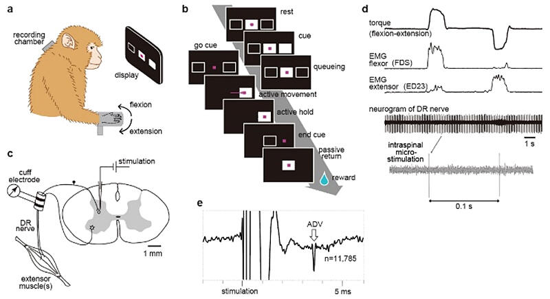
Figure 1: Experimental setup and examples of antidromic volleys.

Provided by NCNP
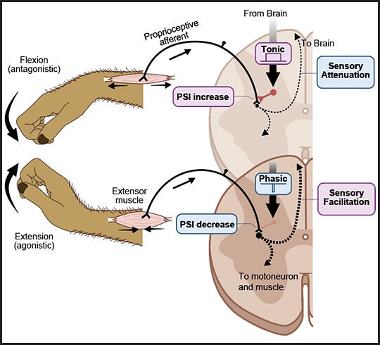
Figure 2: A circuit model for gain modulation of proprioceptive afferent signals by presynaptic inhibition at the spinal cord during agonistic and antagonistic movement.

Provided by NCNP
The mechanism by which the brain processes vast amounts of sensory signals while executing physical movement has, to date, been unclear. In this study, the group weakly stimulated the sensory nerve endings of the wrist extensor muscles in the spinal cord of a monkey during exercise and measured the antidromic potentials (ADVs) from the sensory nerve bundles of the wrist extensor muscles. When the ADV was large, the presynaptic inhibition was also large. The researchers also analyzed how the magnitude of ADV induced by spinal cord stimulation changed during movement.
The researchers discovered that the magnitude of presynaptic inhibition is not constant during exercise but rather changes with the phase of movement. Presynaptic inhibition alters the signal transmission from muscle sensory nerves to the spinal cord depending on the phase of movement.
Furthermore, the researchers' detailed analysis of the temporal changes in ADV revealed that the decrease in presynaptic inhibition during wrist extension began with the onset of muscle activity and was unrelated to the start timing of wrist motion. The observed changes in presynaptic inhibition were caused by motor commands from the brain, equivalent to that producing muscle activity, rather than that from peripheral sensory information caused by movement.
The researchers showed that the regulation of presynaptic inhibition in the spinal cord was controlled by the motor command centers in the brain. Moreover, the brain controls the magnitude of muscle activity by changing the strength of presynaptic inhibition, thereby skillfully controlling wrist movement.
These results are expected to contribute to the clarification of the pathology of many psychiatric and neurological diseases that cause sensorimotor abnormalities, as well as the development of new rehabilitation techniques and motor skill teaching methods in physical education/sports settings.
Seki said, "The peripheral nerve retrograde potential signal used as an indicator of presynaptic inhibition in our research was only one microvolt. It was very difficult for us to develop an experimental technique to record the weak signal alone from various electrical noises in the monkey during voluntary movements. By combining the role of cutaneous nerves, which we published a paper on in 2003, with the presynaptic inhibition for proprioception in this paper, we could understand the role of presynaptic inhibition on somatosensation during exercise."
This article has been translated by JST with permission from The Science News Ltd. (https://sci-news.co.jp/). Unauthorized reproduction of the article and photographs is prohibited.


