A research group led by Specially Appointed Assistant Professor Akihiko Adachi of the Graduate School of Medicine at Chiba University and Dr. Yuri Kawashima of the Graduate School of Arts and Sciences at the University of Tokyo (currently Specially Appointed Assistant Professor of the Research Institute for Radiation Biology and Medicine at Hiroshima University) recorded a video of a phenomenon called Pseudoleukocoria (false white pupil), in which even the pupil of a healthy child appears white, and explains its mechanism and shows the phenomenon to the public for the first time in the world.
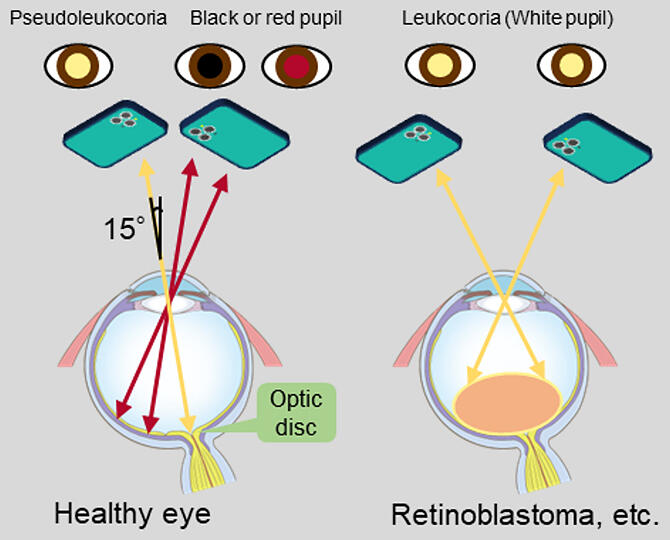
The research group demonstrated that when light enters the pupil at a specific angle at the time of imaging, 'pseudoleukocoria' appears as the reflection from the optic disc. When a white pupil (leukocoria) is observed in a child, parents should take the child to a doctor promptly (as it could present as a sign of a serious medical conditions) but without feeling excessive concern (since it may be a false positive phenomenon, as depicted in this report). The findings were published in the medical journal Pediatrics International.

Provided by Chiba University
Leukocoria' is the symptom of a whitish (yellow or beige) reflection of light from an ophthalmic lesion or fundus structure through the pupil when flash photographs or videos are taken and is a sign of potentially serious conditions including a congenital cataract, retinoblastoma, retinal detachment, persistent hyperplastic primary vitreous, and Coats' disease. In particular, as retinoblastomas are characterized by rapid growth, children showing leukocoria in photographs should visit a specialized medical institution within one week.
Due to the widespread use of smartphones, 'leukocoria' may be noticed more and more by families in digital images. However, white pupils may also be observed in images of healthy children. This phenomenon is called pseudoleukocoria. However, previous medical books and articles illustrate 'pseudoleukocoria' using still images, which limits their interpretability.
In the current study, the research group described 'pseudoleukocoria' using a video to publicize and promote an increased understanding of the difference between a healthy 'pseudoleukocoria' and a dangerous 'leukocoria.' A video can be downloaded from the journal website (https://doi.org/10.1111/ped.15600).
The video capturing 'pseudoleukocoria' consists of 15 frames, with each frame lasting approximately 33ms. 'Leukocoria' caused by retinoblastoma appears as a white reflection from the growing tumor, whereas 'pseudoleukocoria' appears as a white reflection from the optic disc seen in images taken from 15° outward. As 'pseudoleukocoria' is characterized by the reflection of light from the optic disc corresponding to the blind spot, which is a small area, the color of the eye in the video returns to the normal black when the angle of light entering the eye and/or the angle of view is changed slightly.

Provided by Chiba University
Adachi commented, "Dr. Kawashima is a basic researcher of DNA recombination, and I am a neurosurgeon. We published this article on pediatric ophthalmology by pure happenstance. I would like to continue publishing reports on basic and clinical research according to our findings, even those beyond our area of specialty."
Journal Information
Publication: Pediatrics International
Title: Pseudoleukocoria
DOI: 10.1111/ped.15600
This article has been translated by JST with permission from The Science News Ltd. (https://sci-news.co.jp/). Unauthorized reproduction of the article and photographs is prohibited.


