
The research group including Associate Professor Kenbun Sone, Graduate Student Yusuke Toyohara, and Professor Yutaka Osuga of the Graduate School of Medicine at the University of Tokyo, Assistant Professor Ryo Kurokawa of the Department of Radiology at Tokyo University Hospital, and Katsuhiko Noda and Kaname Yoshida of SIOS Technology, Inc. successfully automated the image selection process for AI learning that doctors use to improve diagnostic accuracy and newly developed AI to enable the automated diagnosis of uterine sarcoma. This was published as an Online First Article prior to publication in the Journal of Gynecologic Oncology.
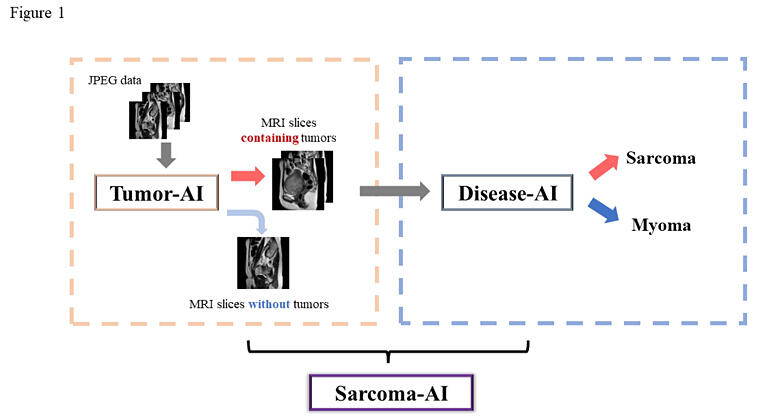
AI, artificial intelligence; MRI, magnetic resonance imaging.
Provided by the University of Tokyo
Uterine sarcoma is a rare cancer with a poor prognosis; in some cases, it is challenging to distinguish it from uterine fibroids with degeneration. Since the treatment strategy differs between uterine sarcoma and uterine myoma, an accurate preoperative diagnosis is necessary. Magnetic resonance imaging (MRI) is considered useful for its identification. The research group used preoperative MRI images of uterine sarcomas and uterine fibroids (263 cases in total) for deep learning and evaluation. The resulting accuracy rate of the automatic uterine sarcoma diagnosis made by AI of the dataset used for cross-validation was 89.32%. The evaluation of an unknown data set (32 cases in total) that was not used for cross-validation yielded a diagnostic accuracy rate of 92.44%.
For the AI to make a diagnosis, a doctor must select only images that include the lesion site, which has been a major challenge for social implementation. Automation has made it possible to input all MRI images obtained in clinical practice, rather than just those of uterine sarcomas and uterine fibroids, as-is into the AI. Future applications of this technology in clinical practice are expected.
Journal Information
Publication: Journal of Gynecologic Oncology
Title: The automatic diagnosis artificial intelligence system for preoperative magnetic resonance imaging of uterine sarcoma
DOI: 10.3802/jgo.2024.35.e24
This article has been translated by JST with permission from The Science News Ltd. (https://sci-news.co.jp/). Unauthorized reproduction of the article and photographs is prohibited.