
A research group led by Professor Shuichiro Inoue of the Institute of Quantum Science at Nihon University and the Institute for Advanced Medical Research of the Keio University School of Medicine announced the development of an ultra-sensitive optical tomography imaging method by applying a quantum technology called quantum pulse gate (QPG) to the time-resolved measurement of light pulses. This imaging method enables non-contact, non-invasive visualization of the depths of a living body through weak-light irradiation and is expected to serve as a novel medical imaging device for medical diagnosis and other purposes. The results were published in the international journal Scientific Reports on November 29, 2023.
Provided by Nihon University
Optical coherence tomography (OCT) is a technique that irradiates a sample with low-interference light and acquires a tomographic image through interference between the reflected light from within the sample and a reference light. It is needed in various fields. In this study, the research group made time-resolved measurements of reflected light from within the sample without using interference with reference light as done in OCT to obtain information on fault structures. They used sum-frequency generation (SFG) to detect light pulses.
SFG converts the frequency of reflected light pulses to a higher frequency through a second-order nonlinear optical process. Simultaneously injecting reflected and pump light pulses into a nonlinear optical crystal generates a third higher-frequency light pulse. This occurs due to the coupling of the two pulses.
The information regarding where the detected light pulses reflected inside the sample is obtained from the time delay of the pump light pulses. A tomographic image of the sample can then be acquired from this information, along with the detected light intensity (photon count).
To achieve high image quality, QPG was used to remove light pulses reflected once inside a living body and returned (signals) and light pulses that arrive simultaneously and undergo scattering and return (background noise).
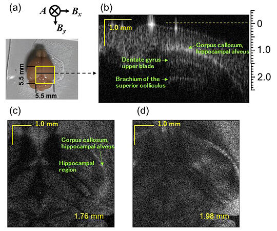
Using a single-photon detector to detect the QPG output, the research group achieved a signal-detection sensitivity that far surpasses that of OCT. Using a mouse brain as the biological sample, they succeeded in visualizing the depth of a living body. A signal detection sensitivity of 111 dB was achieved at a probe-light intensity of 1.5 mW, demonstrating a depth of coverage equivalent to that of OCT using a 1700-nm band SC light as a light source.
Inoue said, "We have been developing single-photon detectors in the optical communication wavelength band and conducting experiments applying them to quantum communications. Since it was our first experiment dealing with biological samples, it was truly exciting when we successfully detected reflected light from inside a mouse brain. Moving forward, we aim to perform tomographic imaging of the living Alzheimer's disease-model mouse brain, enabling the temporal observation of the accumulation of amyloid β, one of the causative factors of Alzheimer's disease. Through this, we hope to unravel the mechanism of dementia onset and anticipate the development of new therapeutic approaches and drugs."
Journal Information
Publication: Scientific Reports
Title: Quantum optical tomography based on time-resolved and mode-selective single-photon detection by femtosecond up-conversion
DOI: 10.1038/s41598-023-48270-7
This article has been translated by JST with permission from The Science News Ltd. (https://sci-news.co.jp/). Unauthorized reproduction of the article and photographs is prohibited.