
Professor Hirokazu Kaji and Assistant Professor Takeshi Hori of the Institute of Biomaterials and Bioengineering at Tokyo Medical and Dental University), in collaboration with Professor Emeritus Takahiro Arima of the Environment and Genome Research Center at the Graduate School of Medicine at Tohoku University, Professor Hiroaki Okae of the Institute of Molecular Embryology and Genetics at Kumamoto University and others, announced that they have succeeded in using human trophoblast stem (TS) cells to generate placental organoids (miniature tissues) that mimic the villous structure of the human placenta. On the basis of these placental organoids, the research group succeeded in developing a placental barrier model with a sheet-like structure. Using this model, substance transfer from a mother to a fetus can be evaluated quantitatively, similar to an evaluation system using the actual placenta. The model is expected to be applied to the assessment of viral, bacterial, and drug influences on fetuses. The results were published in the international academic journal Nature Communications on February 8.
Credit: Department of Diagnostic and Therapeutic Systems Engineering, TMDU
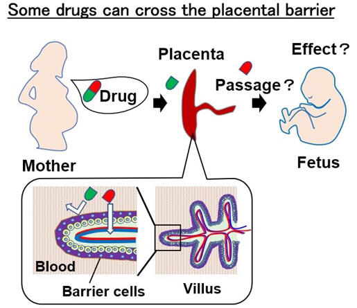
The human placenta serves as a barrier, protecting the fetus from exposure to foreign substances such as pharmaceuticals and viruses. However, some substances can still cross through the placenta and affect the fetus. The human placenta has internal villi. Barrier cells on the villus surface serve the role of sorting out substances (including pharmaceuticals) to be transferred to the fetus. To develop pharmaceuticals with minimal risk to a fetus, a quantitative assessment of the amount of substances transferred is required.
Such assessment using animal experiments is challenging owing to the interspecies variability in placental structure. The complicated structure of the assessment system using actual human placentas and the difficulty in securing a supply are further obstacles. Moreover, as previously developed placental cell culture models were derived from choriocarcinoma cells, accurate assessment was not possible.
In 2018, a research group at Tohoku University succeeded in establishing human TS cells. They confirmed that undifferentiated TS cells can be maintained in a stable state for a long period and are able to differentiate into syncytiotrophoblast cells (barrier cells) that are responsible for hormone production and extravillous trophoblast (EVT) cells that possess infiltration ability. The problem of culturing by means of barrier cells alone is that stable culturing for a long period is difficult.
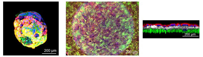
In the current study, the research group considered the development of organoids in which barrier cells and underlying cytotrophoblast cells coexist in the same way as they do in the actual placenta. By exploring the culture conditions, the researchers confirmed that spherical organoids could be generated in culture based on agarose and three types of media. They succeeded in generating placental organoids, on whose surface barrier cells exist and which is filled with undifferentiated cells on the inside.
The developed human placental organoids were confirmed to secrete human chorionic gonadotropin, which is necessary for the continuation of pregnancy, and to possess the unique fusion ability of barrier cells. Additionally, electron microscopy analysis confirmed that the microvilli on the organoids were similar in shape to those on the actual placenta surface.
However, quantitative assessment of compounds was difficult with these spherical organoids.
Therefore, by applying the conditions for generating human placental organoids and using insert column-type well plates, the research group considered the development of a placental barrier model in which the organoids are arranged in sheets. The well plate had a double structure, in which a column was installed in the hollow part of the plate, and the sheet-like placental barrier model was cultured at the bottom of the column. To enable quantitative assessment of substance permeability, the solution to be evaluated was placed in the lower plate of the double structure, and the solution that was transferred through the placental barrier model to the upper plate was analyzed.
It was confirmed that sheet-shaped cells were cultured so as to completely cover the column bottom in a triple layer structure of: a barrier cell layer, a TS cell layer, and an endothelial cell layer. The formation of microvilli on the surface of the barrier cells was also confirmed.
A comparison of quantitative assessment functions between the developed placental barrier model and an actual placenta revealed that both gave similar results. Using dextran of different particle sizes, the permeability of the developed model was confirmed to be similar to that of an actual placenta, and this was further verified by the high permeability of antipyrine (a type of analgesic) and caffeine as well as the low permeability of glyphosate (a type of herbicide).
Aiming for early practical application, the research group will present the developed model to pharmaceutical companies. They will also continue to optimize the form and other aspects of the model. Moreover, they will engage in basic research to clarify which pregnancy term the developed human placental organoids represent.
Credit: Department of Diagnostic and Therapeutic Systems Engineering, TMDU
Hori commented, "The placental barrier model that we developed has potential to be a useful cell culture model for elucidating viral and bacterial influences on a fetus via the placenta, substance transfer to a fetus, and the processes of placenta formation and maturation. Furthermore, the model is expected to contribute to drug discovery and to the development of a new pharmaceutical safety assessment model as an alternative to animal testing."
Journal Information
Publication: Nature Communications
Title: Trophoblast stem cell-based organoid models of the human placental barrier
DOI: 10.1038/s41467-024-45279-y
This article has been translated by JST with permission from The Science News Ltd. (https://sci-news.co.jp/). Unauthorized reproduction of the article and photographs is prohibited.