
A research group led by Graduate Students Shoichiro Yamaguchi and Natsuki Minamide and Lecturer Kazuaki Maruyama, all of the Graduate School of Medicine at Mie University announced that they have clarified the development path of lymphatic vessels form. They accumulated human embryos in residual specimens collected for pathological examination. Various lymphatic markers were used to analyze the process of early lymphatic development and the distribution of lymphatic vessels in various organs. The findings are expected to help shed light on the pathology of various lymphatic-related diseases. The study results were published on February 14 in The EMBO Journal, an international academic journal on molecular biology.
Provided by Mie University
Lymphatic vessels, which are a type of channel such as arteries, are distributed throughout the body. As a critical organ, they are known to be involved in immune functions and edema. They are also found to be distributed in the brain and bones and have been gaining attention as a therapeutic target in recent years.
Based on experiments performed on genetically modified zebrafish and mouse embryos, lymphatic vessels are known to be formed mainly through the transdifferentiation of venous endothelial cells. However, whether the findings in animal models are applicable to humans has been unknown since the genesis and physiological roles of lymphatic vessels differ greatly between species.
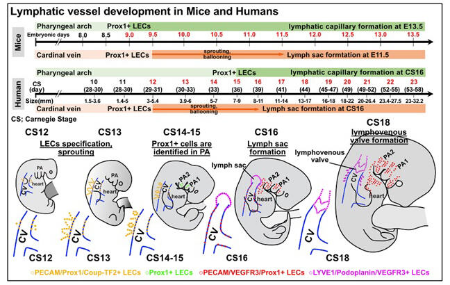
Previous reports on genetic lineage analysis using zebrafish and mouse embryos had revealed that the transdifferentiation of lymphatic endothelial cells (LECs) from venous endothelial cells is mediated by the expression of Prospero homeobox protein 1 (Prox1), a master transcription factor involved in lymphatic vessel genesis. The research group had previously discovered that in the head and neck regions, LECs also originate from undifferentiated mesodermal cells (cardiopharyngeal mesoderm) derived from the first and second pharyngeal arches.
In the current study, they analyzed this discovery further using 3- to 8-week-old human embryos and 9-week-old fetuses. The results showed that LECs within the pharyngeal arches appeared at around Carnegie stage (CS) 14-15. From CS23 to the ninth week, luminal structures were observed in this region. They exhibited very slow genesis, remaining sparse for several weeks before they were formed.
The study also clarified the genesis process of a lymphovenous valve, which is a valve structure that exists on the lymphatic side of the junction between the clavicular vein and internal jugular vein and prevents blood from flowing back into the lymphatic vessel. At CS16, LECs budding from the common cardinal vein formed the large initial lymphatic vessels (lymph sacs). At CS18, protrusions began to appear on the lumen surface at the junction between the lymph sac and the common cardinal vein and gradually transformed into an organized valve structure, which was formed from CS21 to the ninth week. By contrast, during the organogenesis period (3rd to 8th week) and in the 9-week-old fetus, no LECs or lymphatic vessels were observed in the spinal cord or around the brain and parenchyma.
In summary, 1) lymphatic vessels are formed from transdifferentiated venous endothelial cells, as in fish and mice; 2) compared with lymphatic vessels in the trunk, those in the upper body are generated very slowly; 3) at the junction between the initial lymphatic vessel (lymph sac) and the common cardinal vein, a valve that divides the lymphatic vessel and the vein is formed to shape the venous angle; and 4) how lymphatic vessels are to be distributed differently in various organs was clarified.
The study results are expected to help shed light on the pathology of many lymphatic-related diseases, including lymphedema, obesity, cardiovascular disorders, Crohn's disease, and congenital lymphatic malformation.
Maruyama said, "Our study is valuable for translating findings from animal studies to humans. I would like to thank fifth- and fourth-year medical students Yamaguchi and Minamide for their efforts."
Journal Information
Publication: The EMBO Journal
Title: The development of early human lymphatic vessels as characterized by lymphatic endothelial markers
DOI: 10.1038/s44318-024-00045-0
This article has been translated by JST with permission from The Science News Ltd. (https://sci-news.co.jp/). Unauthorized reproduction of the article and photographs is prohibited.