
A group led by Professor Satoshi Yamaguchi, who studies biofunctional chemistry at the Institute of Scientific and Industrial Research at Osaka University, developed a technology for instantly and precisely attaching living cells one by one to a desired location using light irradiation. This technology enabled real-time observation of immune cells attacking cancer cells. They expect that the technology finds applications in a wide range of fields from treatment of cancer and other diseases to industry.
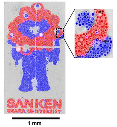
Produced by Yuki Umeda, a second-year doctoral student at the School of Engineering at the University of Tokyo
Previously, cells were trapped in tight holes or sprayed
One reason for recurrence of cancer after anticancer therapy is the heterogeneity of cancer cells. Because the therapeutic efficacy of anticancer drugs varies from a cancer cell to another, some cancer cells are resistant to and survive from drug treatment. To evaluate drug response and other properties of a heterogeneous cell population, it is ideal to use "single cell analysis," in which cells in the population can be observed one by one.
Single cell analysis techniques include confinement of cells in a small space using microscopic holes (microwells) or structures placed in a flow channel (microfluidics); however, shape changes cannot be observed. In the methods of capturing cells with electrodes (dielectrophoresis) and spraying one cell at a time (inkjet printer method), cells adhering to the substrate can be observed; however, floating cells such as leukocytes in blood can flow away during observation and disappear from the field of view.
Switching functionality achieved after 20 years of effort
Yamaguchi began exploring the possibility of using photoreaction-based substrates for single-cell analysis around 2005. Around 2010, during his time working for the University of Tokyo, he synthesized a substrate (first generation) made of glass coated with PEG-lipid, which consists of the water-soluble polymer polyethylene glycol (PEG), a hydrophobic lipid, and a photodegradable molecule (linker) linking the two. Lipids interact with and adhere to the lipid-bilayer membranes of the cells. In the light-irradiated portion of the substrate, however, the linker breaks down and is released, and the PEG portion, which has characteristics that make it difficult to adhere to the cells, appears on the surface layer. It is thereby confirmed that cells are released.
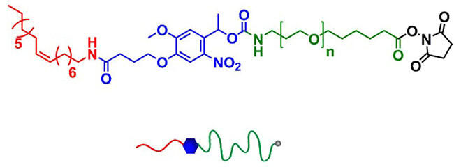
Provided by Professor Satoshi Yamaguchi of Osaka University
Around 2015, Yamaguchi, a former student of his, Program-specific Lecturer Shinya Yamahira, of the Graduate School of Biostudies at Kyoto University, and their research group modified the PEG-lipid to contain two lipid parts. When a linker was attached to one of the lipids and irradiated with light, this version of PEG-lipid (second generation) lost one lipid and could adhere to the cells by the remaining one lipid.
Around 2020, the team changed the linker between PEG and lipid parts from a photodegradable one to a linker that can have a hydrophilic or hydrophobic property depending on the type of light, creating a new version of PEG-lipid with a switching function (third generation), which releases the cells by visible light irradiation and adhere to cells by UV light irradiation.
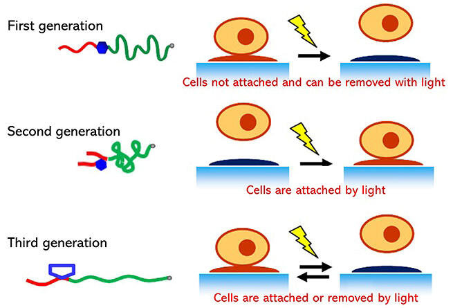
Provided by Professor Satoshi Yamaguchi of Osaka University
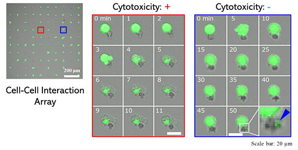
Observing an array of cancer cell-immune cell pairs to understand treatment effects
Using second-generation PEG-lipid, cells that are normally suspended in blood vessels or other locations can be attached to light-exposed areas.
To investigate the heterogeneity of cancer cells, natural killer (NK) cells, which are responsible for immunity, and cancer cells were placed one by one on the array to observe their interaction. It was possible to confirm the process by which an NK cell kills a cancer cell and the process by which an NK cell takes up part of a cancer cell without killing it. This method is expected to contribute to the advancement of cancer treatment.
The desire to create a platform for cell analysis
Aside from medicine, if insect olfactory receptor cells can be attached and detached in any arrangement desired, it is possible to create highly sensitive sensors for specific smell, for example. Yamaguchi enthusiastically stated, "We would like to develop a platform technology that isn't limited to specific fields but is able to contribute to research that requires looking at the phenotype of cells, research and industry in which cells are manipulated in some way and some of the manipulated cells need to be recovered."
In the information industry, GAFAM, a group of five giant IT companies, is attracting attention as a platform. There are expectations that single-cell analysis technology will also become a platform supporting biological research in the future.
(NAGASAKI Midoriko / Science Portal Editorial Office)
Original article was provided by the Science Portal and has been translated by Science Japan.