
A research group led by Assistant Professor Mariko Kikuchi and Professor Minoru Tanaka of the Graduate School of Science at Nagoya University, in collaboration with Hokkaido University and Kyoto University, has announced that they clarified the molecular mechanism by which oocyte polarity (heterogeneity) is created in medaka (Oryzias latipes). They discovered a dome-shaped microtubule structure in germ cells which was found to create oocyte-specific polarity before sex determination, regardless of the sex of the parent. The results are expected to contribute to the elucidation of the mechanisms of germ cell sex determination and the vertebrate body axis formation and were published in the March 13 issue of the international academic journal Development.
Provided by Assistant Professor Mariko Kikuchi and Professor Minoru Tanaka of the Group of Reproductive Biology at Nagoya University
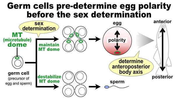
In organisms with different sexes, including in humans, cells must be correctly aligned along the body axis (anterior−posterior, lateral, and vertical axes) to successfully form a vertebrate body from an egg. Among these axes, the anterior−posterior axis is derived from oocyte-specific polarity and creates the anterior−posterior orientation of the body, whereas polarity is not observed in sperm cells.
The sex of medaka is determined by the action of a gene on the Y chromosome on the fourth day after fertilization. Oocytes or sperm cells are created from primordial germ cells or common germ cell precursors, and germ cells are known to be capable of developing into oocytes or sperm before sex determination. Primordial germ cells migrate to the gonads within one day of fertilization, are taken up by the gonads, and then differentiate into germ cells. However, it is unclear how the germ cells acquire the polarity necessary to become oocytes.
The research group has been trying to shed light on the mechanism of sex determination in medaka, which is used as a sexual reproduction model and shares many similarities with humans. Their previous studies have shown that large changes in the expression of genes involved in microtubules coincide with the germ cell commitment to oogenesis or spermatogenesis. In this study, the research group visualized microtubules in germ cells and discovered a new hollow, dome-shaped microtubule structure that occupies a large area inside the cell. They named the structure "microtubule dome." The microtubule domes were present in the parental germ cells before sex determination. After sex determination, microtubule domes were maintained stably if the parent became female, whereas they disappeared if the parent became male.
To investigate the function of this microtubule dome, they examined its involvement with the Balbiani body (an oocyte-specific intracellular aggregate), which was known to be necessary for formation of polarity in the oocyte. The microtubule domes colocalized with the Balbiani bodies, suggesting that they may serve as scaffolds. The Balbiani body was known to be necessary to determine the anterior−posterior orientation of the body.
To verify the relationship between microtubule domes and Balbiani bodies, they disrupted the microtubule domes with reagents and observed the time-dependent dispersion of buckyballs, a component of Balbiani bodies. These findings demonstrated the possibility that microtubule domes create the polarity of future eggs. Subsequently, they examined when the microtubule domes were formed, and found that they were formed in the germ cells in the gonads two days after fertilization, before sex determination.
Since sex is determined four days after fertilization, it indicates that the germ cells are prepared to produce eggs regardless of which sex the parent is. To clarify the molecular mechanism by which the microtubule dome is maintained specifically in females, they examined its relationship with the known oogenesis factor Foxl3. In ovaries of female medaka with mutations in the foxl3 gene, eggs were not produced, and germ cells differentiated into sperm. Unexpectedly, the microtubule dome was maintained stably even in ovaries where the foxl3 gene is not functional. Conversely, to investigate the cause of microtubule dome instability in males, they examined the involvement with dmrt1, which is expressed specifically in somatic cells surrounding male gonadal germ cells. The results showed that dmrt1 of somatic cells destabilizes the microtubule dome structure.
The result indicates that in males, signals from these somatic cells destabilize the microtubule domes, masculinizing the germ cells. These data revealed that the microtubule structure characteristic of oocytes in the gonads is formed in the germ cells before sex determination of the parent, and the germ cells themselves are intrinsically female. The interaction between germ cells and somatic cells was also shown to be important for germ cell masculinization.
Kikuchi said, "Moving forward, we would like to study what effects the microtubule dome structure has on embryonic development, and also advance our research on sex and reproduction from the perspective of the role of somatic cells that control germ cell sex determination."
Journal Information
Publication: Development
Title: Sexually dimorphic dynamics of the microtubule network in medaka (Oryzias latipes) germ cells
DOI: 10.1242/dev.201840
This article has been translated by JST with permission from The Science News Ltd. (https://sci-news.co.jp/). Unauthorized reproduction of the article and photographs is prohibited.