
A research group led by Researcher Yuko Ohnishi and Professor Shimpei Gotoh of the Department of Clinical Application at the Center for iPS Cell Research and Application (CiRA) at Kyoto University, announced their research results, showing that activation of YAP/TAZ signaling and inhibition of AKT signaling promote the differentiation of alveolar type II epithelial cells (AT2) into alveolar type I epithelial cells (AT1), which are responsible for alveolar repair mechanisms after injury due to viral infection and other causes. They established a human iPS cell line that can be used for highly sensitive quantification of AT1 differentiation. The discovery was made using alveolar epithelial cells derived from this cell line to create alveolar spheroids suitable for screening a large number of compounds. The results are expected to be useful for drug discovery for a wide range of lung diseases and were published in the March 28 issue of the international academic journal Stem Cell Reports.
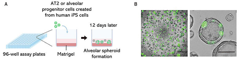
B) Live-cell imaging of on-gel culture of alveolar spheroids derived from GFP+ iAT2 cells. Left scale bar, 500 µm; right scale bar, 100 µm.
Provided by Kyoto University
The human lungs consist of airways and alveoli, and the alveoli are responsible for gas exchange. The alveolar surface is covered with two different types of epithelial cells, namely, AT1 and AT2. AT1 cells are flat and allow oxygen and carbon dioxide to move back and forth. AT2 cells are cuboidal and secrete components that maintain the alveoli's pouch-shaped structure. Typically, when AT1 is damaged due to viral infection or other factors, AT2 cells proliferate as tissue stem cells and some of them differentiate into AT1. In contrast, certain conditions such as interstitial pneumonia and novel coronavirus diseases have been reported to interfere with AT2-to-AT1 cell differentiation, resulting in the formation of abnormal cells.
Meanwhile, the regulatory mechanism of AT2-to-AT1 cell differentiation remained unclear due to the difficulty in obtaining alveolar epithelial cells from humans. Previous studies of the research group aimed to understand lung diseases and develop treatments. They succeeded in generating alveolar organoids from human iPS cells in 2014 and alveolar organoids that can be cultured for a long term in 2017. In 2021, they revealed some of the signals involved in AT1 differentiation.
In this study, the research group aimed at comprehensive screening of signals involved in AT1 differentiation. First, for high-sensitivity and quantitative evaluation of differentiation into AT1 cells, they established iPS cells via gene transfer to visualize AT1 differentiation.
Next, they examined the preparation of alveolar spheroids (spherical cell aggregates), which are easy to prepare and can be used for efficient screening. They developed the "on-gel culture method" for simple formation of alveolar spheroids using a small number of cells. Using this method, alveolar spheroids can be easily formed on the gel by dispensing a suspension of alveolar epithelium to a 96-well culture plate, of which the well bottom is precoated with Matrigel (culture substrate). Moreover, this culture method was used to culture alveolar epithelial progenitor cells induced and differentiated from iPS cells, in which differentiation into AT1 can be visualized. Each of the 274 low-molecular-weight compounds was added to each well, and the effect of promoting differentiation into AT1 was evaluated via luminescence intensity. Compounds that caused extremely reduced cell counts were excluded as they are likely to be cytotoxic.
As a result, 15 compounds produced high luminescence intensities. Of these, five compounds were newly found to increase the expression of three AT1 marker genes. The expression levels of AT1 marker genes were examined, and a YAP/TAZ signal activator (LATS-IN-1) showed particularly increased expression. Elevation of the YAP/TAZ signal has been reported to be important for AT1 differentiation in mice. They also examined the results of simultaneous addition of the YAP/TAZ signal activator and each of the remaining four compounds and found that the AKT signal inhibitor greatly increased the expression of the AT1 marker genes. Increased AT1 marker proteins and genes were observed. Treatment with the two compounds increased AT1 marker-positive spheroids to 90% from 40% without treatment. The effects of the two compounds were also confirmed in AT2 cells collected from humans. Finally, RNA sequencing analysis was performed to comprehensively examine the changes caused by the addition of the two compounds.
The results showed that each compound alone increased the expression of many AT1 marker genes, but the simultaneous addition of the two compounds further enhanced the expression of these genes. Simultaneous addition of both compounds increased angiogenic potential and induced cells with a shape similar to that of AT1 cells. The screening method developed in this study can be automated in the future, and the research team hopes that it contributes to more efficient drug candidate discovery and regenerative medicine.
This article has been translated by JST with permission from The Science News Ltd. (https://sci-news.co.jp/). Unauthorized reproduction of the article and photographs is prohibited.