
A research group led by Professor Masayuki Masu of the Institute of Medicine at the University of Tsukuba and Specially Appointed Assistant Professor Seiichi Koike of the Graduate School of Science and Engineering at the University of Toyama, in collaboration with Yokohama City University, the National Institutes of Natural Sciences' Exploratory Research Center on Life and Living Systems, and the National Institute for Physiological Sciences, has announced that they developed a technique using fluorescent substances to label the vesicles, which are involved in intracellular transport of substances and other functions, and visualize the fusion of the labeled vesicles using cells in the yolk sac surrounding a mouse embryo. Observations of the vesicle fusion process revealed two distinct modes of fusion and involvement of cytoskeletal actin in the vesicle fusion regulation. The results are expected to contribute to the elucidation of the regulatory mechanisms of vesicle fusion and trafficking and were published in the international academic journal eLife on March 18.
Provided by the University of Tsukuba
Cells use nutrients and other substances taken up from the outside via a process known as endocytosis. The incorporated substances are encased in endosomes, which are sac-like structures responsible for sorting substances and transported to organelles. Intracellular vesicles fuse with each other or with other organelles during vesicle trafficking. The individual fusion processes are too minute to observe under a microscope, and the mechanisms that control fusion remained unknown.
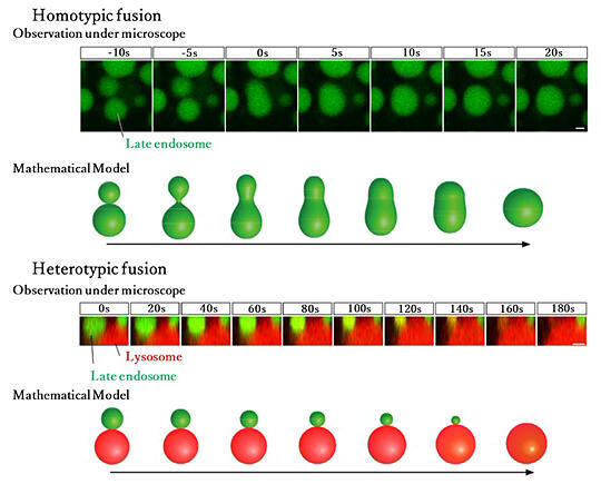
With this in mind the research group used the technology they developed here to observe living mouse embryos at an early stage of development (around embryonic day 8.5). Through this they successfully visualized endosome fusion in the yolk sac visceral endoderm cells, which are in the most superficial portion of the mouse embryo. The results of this observation revealed two distinct modes of endosome fusion: "homotypic fusion," in which two endosomes fuse rapidly to form a single vesicle, and "heterotypic fusion," in which endosomes are slowly absorbed by lysosomes.
Mathematical analysis of the vesicle fusion process using a dynamic model indicated that the fusion mode primarily depends on the vesicle size, with homotypic fusion occurring when vesicles are smaller and heterotypic fusion when vesicles are larger. They also confirmed that large vesicles undergo homotypic fusion when fluctuation forces are applied to the membrane of the vesicles.
Cytoskeletal proteins called actin are bound to endosomes, and actin seems to generate fluctuation forces that promote homotypic fusion. Moreover, cofilin (a protein that severs and depolymerizes actin filaments), which promotes actin turnover, and myosin (a motor protein that moves on actin), which interacts with actin, were also found to be important for vesicle fusion. Masu said, "This study has its origins in the discovery of abnormalities in vesicle trafficking in yolk sac visceral endodermal cells when studying mice dying during early embryonic development. In pursuing the question of why vesicle trafficking was abnormal, we found that late endosome fusion occurs in two modes, and that the fusion modes are regulated by a cytoskeletal protein called actin. Then, we performed mathematical analysis of vesicle fusion and discovered that actin was providing the force to induce homotypic fusion in large vesicles. Vesicle trafficking is a fundamental property of cells, but it has been recently associated with Alzheimer's disease. The results of this study may contribute to the diagnosis and treatment of the disease."
Journal Information
Publication: eLife
Title: Actin dynamics switches two distinct modes of endosomal fusion in yolk sac visceral endoderm cells
DOI: 10.7554/eLife.95999
This article has been translated by JST with permission from The Science News Ltd. (https://sci-news.co.jp/). Unauthorized reproduction of the article and photographs is prohibited.