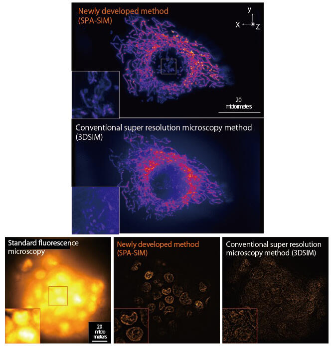
Among super-resolution microscopy techniques suitable for the observation of high-resolution samples, structured illumination microscopy is used for the observation of biological structures and movements because it allows for high-speed observation under relatively weak light. However, observation of the inside of living organisms is currently limited to the near surface layer or thin bodies because background light from outside the focal region of the objective lens hampers observation.
A research group led by Professor Katsumasa Fujita of the Graduate School of Engineering at Osaka University has developed a new super-resolution microscopy method called "selective plane activation-structured illumination microscopy (SPA-SIM)," which enables the observation of the insides of living organisms.
This method uses light-sheet illumination thinner than the focal point of the objective lens and reversibly photoswitchable fluorescent proteins to limit light emission from the sample to the plane observed, thus suppressing background light. By illuminating the object with stripes of light and detecting fluorescence, the insides of thick cells or three-dimensional (3D) multicellular tissue consisting of multiple cells can be observed. The method can also be applied to high-resolution observation of the internal structures of "spheroids," which are clusters of multiple types of cells that are difficult to observe using conventional methods. The spatial resolution was as high as 140 nm (nanometer is one billionth of a meter) in the in-plane direction and 300 nm in the depth direction.
Recently, 3D multicellular tissues such as organoids and spheroids, which are known as mini organs, have been attracting attention in research fields such as biological development and etiology. The new method developed in this study allows the observation of the internal structures of single cells in living organisms and the spatial distribution of multicellular tissues, and it is expected to contribute to biological and pharmaceutical research.


