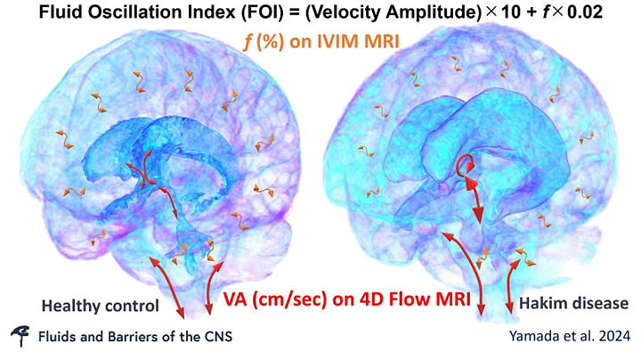
A joint research group led by Lecturer Shigeki Yamada of the Department of Neurosurgery at Nagoya City University Graduate School of Medical Science, together with Shiga University of Medical Science, the University of Tokyo, Osaka University, Tokyo Institute of Technology, Tokyo Metropolitan University, Tohoku University, Yamagata University, and Fujifilm has announced that they have developed a method to observe human cerebral circulation and cerebrospinal fluid dynamics macroscopically using computer simulations. This was achieved by integrating the bidirectional reciprocal motion of cerebrospinal fluid obtained via four-dimensional flow MRI and IVIM MRI. Using this method, they succeeded in clarifying the mechanism of abnormality of cerebrospinal fluid dynamics in Hakim's disease (idiopathic normal-pressure hydrocephalus, iNPH). The findings are expected to contribute to the elucidation of the mechanisms underlying aging and diseases of the brain. The results were published on May 30 in Fluids and Barriers of the CNS, the official journal of the International Society for Hydrocephalus and Cerebrospinal Fluid Disorders.

Provided by Nagoya City University
Cerebrospinal fluid regulates pressure by reciprocating movements through the cerebral aqueduct and subarachnoid space of the foramen magnum in accordance with brain pulsations, allowing smooth blood flow to the brain. Various magnetic resonance imaging (MRI) methods have been developed and are clinically used to observe fluids flowing in the human body without the use of contrast media. Of these, four-dimensional flow MRI and IVIM MRI have been known to provide data that can be used to quantify the velocity components through analysis. Four-dimensional flow MRI captures the flow velocity of fluids (blood and cerebrospinal fluid) in the body during a single heartbeat as tri-axis phase images (in anterior-posterior, vertical, and lateral directions), which can be integrated to observe three-dimensional fluid motion. IVIM MRI can separate and present perfusion and constant directional movement indicative of microcirculation and the random movement and free diffusion of water molecules.
The research group developed a method to quantitatively observe the complex dynamics of cerebrospinal fluid using these two techniques. They had previously reported that fast, complex reciprocal movements of cerebrospinal fluid could be observed using 4D flow MRI, whereas minute slow movements that were not captured could be observed with f values calculated with IVIM MRI. In this study, they examined the feasibility of modeling the movement of human cerebrospinal fluid in the entire intracranial space. The integration of the two methods was considered. First, in 127 healthy volunteers aged 20 years or older and 44 patients with Hakim's disease, 4D flow MRI for cerebrospinal fluid observation and diffusion-weighted MRI with six b values of 0, 50, 100, 250, 500, and 1000 s/mm2 were performed on a high-resolution 3-Tesla MRI scanner. The reciprocal movement of cerebrospinal fluid was observed using the "4D Flow" and "IVIM Analysis" applications on Fujifilm's "SYNAPSE VINCENT" workstation. As a result, the f-value on IVIM MRI corresponding to a flow velocity amplitude of 0.4 cm/s was found to be 75%. Subsequently, they used the IVIM MRI f-values measured in the entire intracranial space to estimate the flow velocity amplitude and introduced a new index, FOI, by combining the two parameters. They successfully developed a method for the macroscopic observation of cerebrospinal fluid dynamics in the entire intracranial space.
With this method, they revealed age-related changes in reciprocal cerebrospinal fluid movement in healthy participants and pathological cerebrospinal fluid movement in patients with Hakim's disease, which is common in elderly people aged 60 years or older. While even healthy individuals aged 60 years or older have enlarged ventricles and increased reciprocal cerebrospinal fluid movement through the cerebral aqueduct, this reciprocal movement is more intense in patients with Hakim's disease. In Hakim's disease, the parietal region of cerebrum and the subarachnoid space are compressed because of the enlargement of the ventricles and Sylvian fissure, resulting in reduced brain pulsations and smaller reciprocal cerebrospinal fluid movements in the lateral ventricles and a large part of the subarachnoid space.
Yamada said, "The ventricles increase in size in some people aged 60 years or older. When the ventricles enlarge beyond a certain size, the subarachnoid space around the base of the skull, called the basilar cistern, also enlarges, pushing the brain upward and reducing the size of the convex part of the subarachnoid space. These changes are considered to underlie the characteristic finding of disproportionately enlarged subarachnoid space hydrocephalus (DESH) and the development of Hakim's disease with symptoms such as gait disturbance, cognitive impairment, and urinary incontinence. However, the mechanism of DESH is not fully understood. In this study, we macroscopically observed the cerebrospinal fluid dynamics across the entire intracranial space."
Journal Information
Publication: Fluids and Barriers of the CNS
Title: Modeling cerebrospinal fluid dynamics across the entire intracranial space through integration of four-dimensional flow and intravoxel incoherent motion magnetic resonance imaging
DOI: 10.1186/s12987-024-00552-6
This article has been translated by JST with permission from The Science News Ltd. (https://sci-news.co.jp/). Unauthorized reproduction of the article and photographs is prohibited.


