A research team led by Special Postdoctoral Researcher Osamu Takenouchi, Special Postdoctoral Researcher Yogo Sakakibara (at the time of research), and Team Leader Tomoya Kitajima of the Laboratory for Chromosome Segregation at the RIKEN Center for Biosystems Dynamics Research has clarified why smaller chromosomes are more prone to aneuploidy in aged oocytes using a new technique to track individual chromosomes in dividing cells. The results were published in Science. Kitajima said, "With the new dynamic imaging technique we developed, we were able to see the movement of individual chromosomes for the first time. We hope to apply this technique to clarify the mechanism behind embryonic development in fertilized eggs."

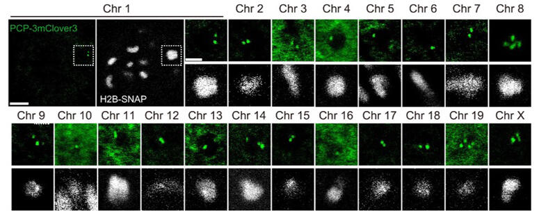
Visualization of target chromosomes in mouse oocytes. Fluorescence signals were detected 5 hours after GVBD. z-confocal sections of H2B-SNAP (chromosomes, white) and PCP-3mClover (target repeat sequence, green) are shown (scale bar, 5 µm). Chromosomes with fluorescent spots are magnified (scale bar, 3 µm).
Provided by RIKEN
The research team has previously analyzed chromosome segregation errors in mouse oocytes and found that the premature separation of homologous chromosome pairs (bivalent chromosomes) during meiosis in aged oocytes is the cause of chromosome segregation errors. However, with conventional live imaging techniques, chromosomes cannot be identified, and it has not been possible to determine the process by which specific smaller chromosomes undergo segregation errors.
In response, they applied CRISPR-Sirius, which fluorescently labels DNA sequences in viable cells, to mouse oocytes to fluorescently label the DNA sequences found only in one type of chromosome, expecting the chromosomes with fluorescent spots to be identified as the target chromosomes. Based on the mouse genome information, 182,147 DNA sequences were selected as candidate labeling sites, prioritized based on accuracy and other factors, and labeled one by one through CRISPR-Sirius. By selecting DNA sequences showing strong fluorescent spots, they succeeded in developing a fluorescent probe set for identifying all 20 chromosomes (19 autosomes and X chromosome) in mouse oocytes. Combined with the microscopic technique they developed for automated location tracking and imaging of chromosomes during meiosis, this new chromosome analysis technique was named the "chromosome tracking-and-identification" method.
Using this technique, they analyzed the dynamic behavior of each of the nine chromosomes (larger chromosomes: chromosomes 1−3 and X, medium-sized chromosomes: chromosomes 8−9, and smaller chromosomes: chromosomes 17−19) in oocytes from young mice. The results showed that smaller chromosomes prefer to move toward the inner region of the spindle during prometaphase, while larger chromosomes prefer to move toward the outer region of the spindle. The results also revealed that smaller chromosomes tend to stretch earlier along the spindle axis. These findings suggest that oocytes regulate chromosome dynamics by altering the timing of chromosome stretching according to size. Smaller chromosomes also preferred to move toward the inner region of the spindle in aged mouse oocytes, like the observation in young oocytes. However, the smaller chromosomes were separated prematurely during meiosis I at a higher frequency, and 60% of the prematurely separated chromosomes became aberrantly distributed in late meiosis I. Since premature separation of chromosomes is caused by the forces of microtubules pulling chromosomes toward the spindle poles, the forces exerted on chromosomes from microtubules were estimated from the elongation of each chromosome. Chromosomes in the inner region of the spindle were more elongated than those in the outer region. The stronger pulling forces on the chromosomes in the inner region of the spindle induced premature chromosome separation.
Takenouchi commented, "We found that with aging, chromosome adhesion molecules decrease in the concentration and move toward the inner region, whereby they cannot withstand the tension, and smaller chromosomes undergo premature separation."
The frequency of premature separation decreased when the artificial kinetochore beads recently developed by the research team were introduced into aged oocytes to prevent the chromosomes from moving toward the inner region, suggesting that the segregation errors may be controllable with the beads.
Journal Information
Publication: Science
Title: Live chromosome identifying and tracking reveals size-based spatial pathway of meiotic errors in oocytes
DOI: 10.1126/science.adn5529
This article has been translated by JST with permission from The Science News Ltd. (https://sci-news.co.jp/). Unauthorized reproduction of the article and photographs is prohibited.


