A research group led by Professor Kenji Izumi (Division of Biomimetics) and Dentist Eriko Naito (Division of Oral and Maxillofacial Surgery) of the Graduate School of Medical and Dental Sciences at Niigata University, and Associate Professor Kazuyo Igawa (Special-Appointment) of the Neutron Therapy Research Center at Okayama University, in collaboration with Group Leader Takashi Shimokawa and Deputy Director Tsuyoshi Hamano of the National Institutes for Quantum Science and Technology (QST) have jointly clarified the cell biological effects of heavy particle beam (carbon ion beam) irradiation on 3D models of human oral cancer and normal oral mucosa. They confirmed that a 3D model of human oral cancer can be used to evaluate therapeutic heavy-particle beam irradiation. The models are expected to find application in comparisons with other particle beams, evaluation of side effects, and to contribute to clarifying the mechanism of therapeutic effects. The results were published in the August 7 issue of the international journal In Vitro Cellular & Developmental Biology-Animal.

Provided by Niigata University
Compared to X-rays, particle beams are less invasive to the normal tissues surrounding cancer, have milder side effects, and are expected to be applicable to not only patients with refractory cancer but also elderly patients. Until now, cell biological evaluation of cancer irradiation has been performed using laboratory animals implanted with planar culture cells or cancer cells. Meanwhile, the three-dimensional positional relationships of cells were not established in planar culture, and the experimental animals differed significantly from humans in the in vivo cancer tissue environment. The challenge was that neither of the evaluation systems could accurately reproduce human clinical trials.
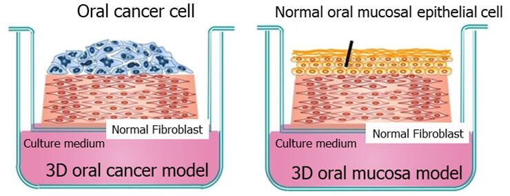
Previously, the research group has been working on the in vitro 3D construction of biological cells to create 3D models useful for the analysis of therapeutic effects and pathological mechanisms. In this study, the research group constructed 3D models of human oral cancer using two types of human oral cancer cells of different grades and a 3D model using normal human oral mucosal epithelial cells. Specifically, fibroblasts were mixed with collagen to construct a cell layer that mimics the submucosal connective tissue of the oral cavity. The 3D models were constructed in 18 days by seeding each type of cells on top of the connective tissue-like layer and continuing incubation while exposing the culture to air during the process of model creation.
A three-dimensional structure in which nutrients are supplied to cancer cells from the undersurface through the underlying cell layer, as in human oral cancer tissue, was reconstructed. The completed models received a single dose of irradiation in a dedicated experimental irradiation room of the heavy-ion cancer therapy device "HIMAC" at QST. The models were taken back to the laboratory, incubation was continued, and observation conducted after 3, 5, and 7 days.
Histopathological observation using optical microscopy, staining with markers related to cell proliferation and death, including apoptosis, quantitative evaluation, and concentration measurement for various proteins secreted into the culture medium were performed.
The results showed that all of the above-listed analytical methods can be used for radiotherapy evaluation. The analytical methods based on the 3D models are also expected to be useful as an alternative method that is more accurate than testing in experimental animals.
Izumi said, "Use of the 3D models we created is expected to allow for comparing heavy particle beams with proton and other particle beams on the same basis, evaluating side effects and their effectiveness in combination with radiation therapy, and studying other aspects of heavy particle beam irradiation. We hope to contribute to the evaluation of cancer radiotherapy by incorporating blood vessels and immune system cells into our models to create more advanced 3D models that are closer to the in vivo environment."
Journal Information
Publication: In Vitro Cellular & Developmental Biology-Animal
Title: The effects of carbon-ion beam irradiation on three-dimensional in vitro models of normal oral mucosa and oral cancer: development of a novel tool to evaluate cancer therapy
DOI: 10.1007/s11626-024-00958-4
This article has been translated by JST with permission from The Science News Ltd. (https://sci-news.co.jp/). Unauthorized reproduction of the article and photographs is prohibited.


