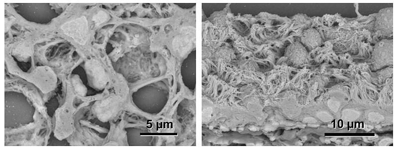
The various organs in the living body have elaborate three-dimensional structures that allow them to perform their physiological functions. Reproducing these is an urgent task in the research and development of regenerated organs. However, the conventional electron staining method developed by Watson in 1958 requires strictly regulated uranium compounds, so its application has been limited to specific research institutions.
A research group led by Professor Akira Sawaguchi of the Faculty of Medicine at the University of Miyazaki established a method for three-dimensional observation of thick sections of biological tissue and cells using an electron microscope by utilizing paraffin for optical microscopy. Electron microscopic imaging takes advantage of the characteristics of low-vacuum scanning electron microscopy to bridge the gap between optical microscopy and electron microscopy. Its wide application to medical and biological research is expected to advance research to elucidate the correlation between biological tissue/cell structure and function. The study was published in npj Imaging.

Provided by the University of Miyazaki
The research group succeeded in developing a simple and rapid method of electron staining using the oxidation reaction of potassium permanganate, achieving a breakthrough that revolutionized ultrastructural analysis of paraffin sections for optical microscopy. The new electron staining method is a simple and rapid protocol in which paraffin sections for optical microscopy are treated with 0.2% potassium permanganate solution for 5 minutes, rinsed with water, treated with Reynold's lead stain for 3 minutes, and rinsed and dried before observation.
Elemental analysis of electron-stained paraffin sections revealed that the amount of lead deposition was enhanced through the unique oxidative action of potassium permanganate and provided the backscattered electrons sufficient to visualize the microstructure of podocytes in the glomerulus and ciliated epithelial cells in the trachea. With this staining method, nano-level ultrastructures that cannot be observed with optical microscopy could be confirmed.
In the published paper, the research group also reported on applying three-dimensional ultrastructure images using the thick section observation method and correlative light and electron microscopy (CLEM) compared with the conventional staining with uranyl and plumbic compounds.
Journal Information
Publication: npj Imaging
Title: KMnO4/Pb staining allows uranium free imaging of tissue architectures in low vacuum scanning electron microscopy
DOI: 10.1038/s44303-024-00045-z
This article has been translated by JST with permission from The Science News Ltd. (https://sci-news.co.jp/). Unauthorized reproduction of the article and photographs is prohibited.


