
A research group including Graduate Student Ikue Shibasaki of the Department of Integrated Applied Life Science, Integrated Graduate School of Medicine, Engineering, and Agricultural Sciences of the University of Yamanashi, Professor Teruhiko Wakayama of the Advanced Biotechnology Center of the University of Yamanashi, and the Konohana Clinic (Kai City, Yamanashi Prefecture) has announced that they succeeded in extracting and analyzing abnormal chromosomes from mouse fertilized eggs with chromosomal abnormalities. Their findings include that about half of the micronuclei contained fragments of two or more different chromosomes and that abnormal chromosome segregation was not associated with specific chromosomes. The findings are expected to contribute to elucidation of the causes of, and development of preventive methods for, abnormal chromosome segregation. The results were published in the international journal Communications Biology on December 26.
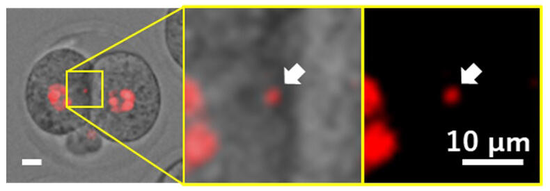
Micronuclei are several micrometers in diameter, so they cannot be seen under bright field conditions, but they can be observed using a fluorescence microscope.
Provided by the University of Yamanashi
Abnormal chromosome segregation in fertilized eggs is one of the causes of miscarriage. Micronuclei formation from abnormal chromosomes occurs at the two-cell stage, but the micronuclei have not been studied in detail because they are small (several micrometers in diameter). Therefore, in fertility clinics, high-grade embryos are selected based on observable indicators such as embryo appearance and developmental rate. However, identification of these embryos in such a manner has been difficult because abnormal embryos with micronuclei are often normal in terms of appearance and early development. Methods that have been used to examine chromosomes with abnormal segregation and micronuclei include comprehensive gene analysis using a single cell in a fertilized egg and observation of chromosomes in formalin-fixed fertilized eggs, but these methods have proven unable to identify abnormal chromosomes.
In this study, the research group developed a technique for extracting micronuclei from mouse two-cell embryos with chromosomal abnormalities without killing the embryos and attempted gene analysis of the extracted micronuclei. To increase the number of embryos with chromosomal abnormalities, freeze-dried mouse sperm were used for intracytoplasmic sperm injection into oocytes, and mRNA for fluorescent staining of chromosomes and other components was injected into the resulting zygotes. Next, to extract the micronuclei from two-cell embryos, they analyzed the relationship between the micronuclei and the cytoskeleton as well as the presence or absence of centromeres on chromosomes and revealed the state of the micronuclei in the two-cell embryos. The micronuclei successfully extracted were subjected to comprehensive gene analysis (DNA-Seq method) one by one to determine the number, structure, and chromosome number of chromosomes found in the micronuclei.
The analysis of the extracted micronuclei revealed that the majority of micronuclei contained only one chromosome, but some micronuclei contained up to four chromosomes. Moreover, any specific chromosomes were found in micronuclei at random frequencies, regardless of chromosome size.
Moving forward, the research group will clarify the normality of fertilized eggs from which abnormal chromosomes have been extracted and the causes of abnormal chromosome segregation, aiming to greatly improve birth outcomes. In addition to extracting the chromosomes, they also succeeded in implanting them into a different egg. In the future, the technology is expected to find application in the normalization of embryos with abnormal chromosome numbers, such as those with Down syndrome and Turner syndrome.
Wakayama said, "Abnormal chromosomes are only a few micrometers in size, so it is extremely difficult to extract them from a fertilized egg with a diameter of 80 micrometers, and no one has succeeded until now. After the long trial and error process, we finally succeeded in extracting and analyzing the abnormal chromosomes without destroying the fertilized egg. Genetic testing is currently used to find and eliminate abnormal fertilized eggs, but we believe that it will become possible to treat fertilized eggs in the future."
Journal Information
Publication: Communications Biology
Title: Extracting and analyzing micronuclei from mouse two-cell embryos fertilized with freeze-dried spermatozoa
DOI: 10.1038/s42003-024-07358-0
This article has been translated by JST with permission from The Science News Ltd. (https://sci-news.co.jp/). Unauthorized reproduction of the article and photographs is prohibited.