
A research team including Professor Takumi Sannomiya of the School of Materials and Chemical Technology at the Institute of Science Tokyo, Dr. Akira Yasuhara of JEOL Ltd., Fumio Hosokawa of FH Electron Optics, Associate Professor Sadayuki Asaoka of Kyoto Institute of Technology, and Professor Tomokazu Iyoda of Doshisha University developed a phase-imaging method that enables uniform information transmission from low to high spatial frequencies by introducing a semicircular aperture in the scanning transmission electron microscope. Because this method allows for easy and inexpensive contrast enhancement, it is expected to advance research and development of organic materials, biological samples, and magnetic materials, for which achieving high contrast is difficult. The results were published online in Ultramicroscopy.
Although electron microscopes enable the visualization of the material structure at a high spatial resolution on the atomic scale, it is difficult to visualize the structures of light elements, such as organic and biological materials, with a size of tens to hundreds of nanometers at high contrast due to weak electron-beam scattering and a low spatial frequency.
Recently, a phase-imaging method using phase plates, which is equivalent to the phase-contrast method employed in optical microscopy, has begun to be used in electron microscopy to solve this problem. However, in the conventional method, the phase plates deteriorate over time due to electron beam irradiation. By introducing a semicircular aperture instead of a circular aperture used in the usual bright-field imaging method of scanning transmission electron microscopy, the researchers developed a phase-imaging method with a phase-contrast-transfer function that includes low and high spatial frequencies. This contrast-transfer function includes a differential phase-contrast component in addition to the usual bright-field image component, resulting in uniform intensity at all spatial frequencies, regardless of focus.
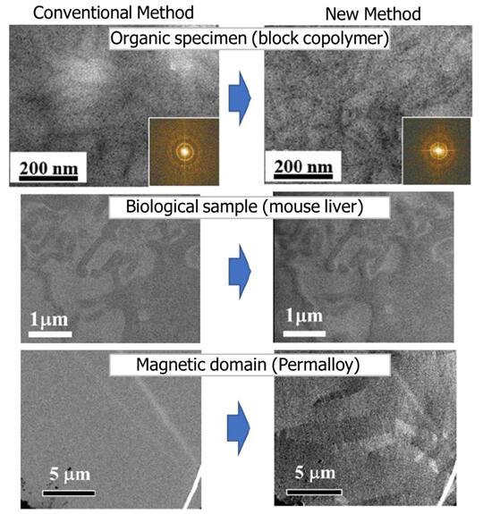
The researchers employed this method for visualizing organic materials, biological samples, and magnetic samples and confirmed that it imaged at higher contrast than the conventional bright-field imaging method. In particular, organic materials and biological samples showed large contrast enhancement at the edges, corresponding to the differential phase-contrast component. For magnetic materials, magnetic domains that could not be observed at all using the conventional method were clearly visualized via the newly developed method.
The newly developed method makes high-contrast phase imaging very simple by introducing a semicircular aperture diaphragm to an ordinary scanning transmission electron microscope. High-quality imaging is thereby enabled with an inexpensive setup that is almost the same as that of an ordinary electron microscope. This method can be used in the biological field to observe biological tissues in their raw state without staining, which has been required in the past. In the case of organic materials, observation after staining has been the norm due to concerns regarding structural changes in the material. However, the newly developed method enables the observation of the material structure as it is and is expected to be applied to material development and analysis in the future. In addition to structural imaging, this method can be used to analyze the distribution of electric potential (electric field) in semiconductors and magnetic field (magnetic domain) in magnetic materials and is expected to be applied to device development in the future.
Journal Information
Publication: Ultramicroscopy
Title: Semicircular-aperture illumination scanning transmission electron microscopy
DOI: 10.1016/j.ultramic.2025.114103
This article has been translated by JST with permission from The Science News Ltd. (https://sci-news.co.jp/). Unauthorized reproduction of the article and photographs is prohibited.