
Recent advances in optogenetic techniques have made it possible to control neuronal activity by exposing the organism to light from the outside. However, conventional optical fiber-based techniques are effective for controlling single neuronal populations but ineffective for manipulating multiple regions simultaneously and precisely. Moreover, light stimulation alone is insufficient to fully understand the complex mechanisms of information processing and propagation in neural networks. Thus, the development of high-resolution techniques capable of measuring the induced neural activity has been awaited.
A research group led by Associate Professor Hiroto Sekiguchi of the Department of Electrical and Electronic Information Engineering and Graduate Student Gota Shinohara of the master's program in Electronics and Information Engineering at Toyohashi University of Technology and Professor Takuya Sasaki and Specially Appointed Researcher Tasuku Kayama of Tohoku University's Graduate School of Pharmaceutical Sciences has developed a hybrid probe integrating micro-LEDs with neural electrodes. The developed hybrid probe enables precisely controlling neural activity in deep biological tissue and simultaneously recording neural activity at multiple points. Using this new hybrid probe, they successfully recorded the induction of specific neural activities by light stimulation in the mouse brain with high spatial-temporal resolution. The new probe can dramatically advance our understanding of neural networks. The study was published online in Applied Physics Express.
Provided by Toyohashi University of Technology
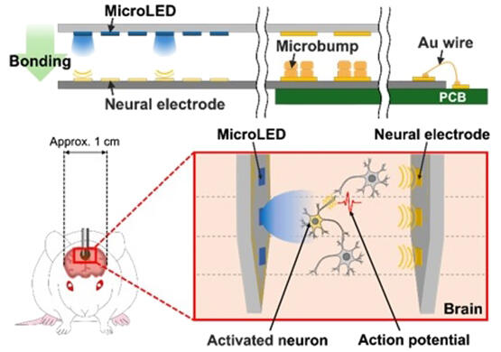
Recently, multi-point light stimulation devices based on micro-LEDs have enabled the individual control of multiple neuronal populations and are expected to significantly enhance our understanding of neural network dynamics. The research group developed a new needle-type hybrid device that integrates micro-LEDs with neural electrodes, enabling light stimulation and simultaneous neural activity recording deep within living tissue.
Micro-LED probes allow for light irradiation to specific sites, and neural electrode probes measure electrical signals of neural activity. The group developed a proprietary technology to manufacture these probes independently, precisely adjust the position of each probe and bond them together precisely with Au microbumps. High spatial resolution was achieved by adjusting the gap between the LED and the electrode in 10-micrometer increments using advanced Au microbump control technology. The angular difference between the bonded probes is 0.02 degrees or less, and their excellent parallel alignment enables smooth insertion into deep regions of the mouse brain.
Furthermore, the research group inserted a hybrid probe with six micro-LEDs and six neural electrodes into the mouse brain, successfully delivering light to a specific area and inducing and recording neural activity in the surrounding region. This hybrid probe overcomes the spatial limitations of conventional optogenetic techniques, allowing for precisely controlling and measuring neural activity. It serves as a powerful tool for understanding neural network dynamics and opens new possibilities for developing therapeutic approaches to neurological diseases. Moving forward, the research group is expected to integrate more advanced multi-point stimulation with recording functions for practical use of the hybrid probes in neuroscience research and medical care.
Journal Information
Publication: Applied Physics Express
Title: Hybrid probe combining MicroLED and neural electrode for precise neural modulation and multi-site recording
DOI: 10.35848/1882-0786/adaf0a
This article has been translated by JST with permission from The Science News Ltd. (https://sci-news.co.jp/). Unauthorized reproduction of the article and photographs is prohibited.